There was a surprising number of new instruments or new versions of existing equipment at this year’s show. As has been the case for some time, most advances attracting attention were in the realm of imaging but there were also some interesting hints at new methods of image transfer and analysis that signal future inroads in the world of telemedicine.
OCT
There was much excitement around the Heidelberg Engineering UK stand as they used the show to launch the new Heidelberg Spirit OCT (figure 1). This is quite a departure for the company known for its Spectralis OCT as the new instrument is aimed fair and square at the primary care optometry clinic.
The instrument is a much streamlined OCT and enables the practitioner to quickly and simply choose from three options (retina, disc and anterior) and offers high resolution capture to meet the demands most likely required in a busy practice.
It is ideal for those wishing to enter the OCT market and requires the minimum of training to capture easily analysed and referable data. Look out for a more detailed review of this soon.
Fans of the Spectralis were also offered a treat in being able to see for the first time a selection of uniquely designed instruments (figures 2 and 3).
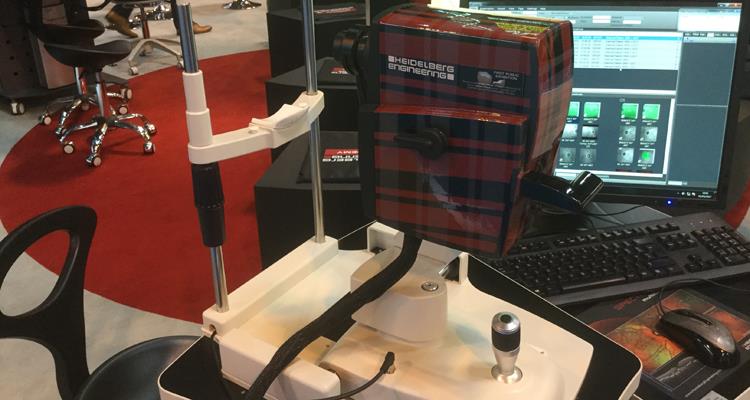
Figure 2
As Emily Malbon, marketing communications manager, explained, ‘The collection comprises custom Spectralis camera casings that were specially made and gifted by the manufacturer, Wild GmbH, to Christoph Schoess, director of Heidelberg Engineering GmbH, personally. The camera casings on show are painted in a variety of unique colours and patterns that mark special occasions in the history of the company. The collection includes a tartan patterned case in tribute of the International Spectralis Symposium 2011 that was held in Edinburgh, which was also the same year Heidelberg Engineering UK was formed. Other colours celebrate the 25th anniversary in 2015 and the opening of the Swiss subsidiary in 2016.’
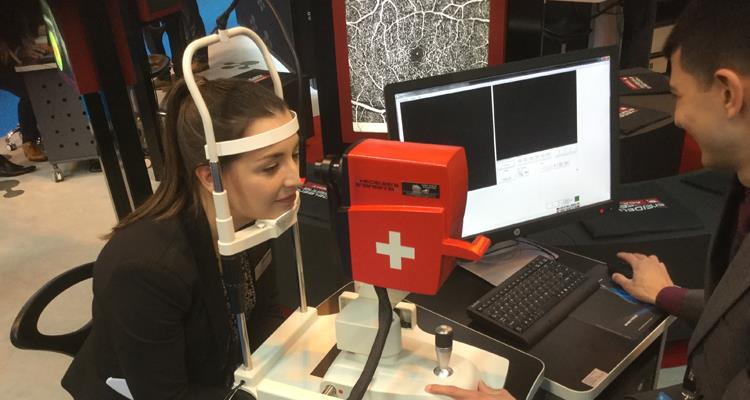
Figure 3
Another new OCT launch was the latest instrument from Optopol found at the BIB Ophthalmic Instuments stand (figure 4 – with Optopol representatives Jaroslaw Wolski and Arkadiusz Chalecki). The new Revo NX has all the operational simplicity we reported on the Revo unit (Optician 24.02.17) but with much faster scanning rates, better resolution and the capability of upgrading to OCT angiography.
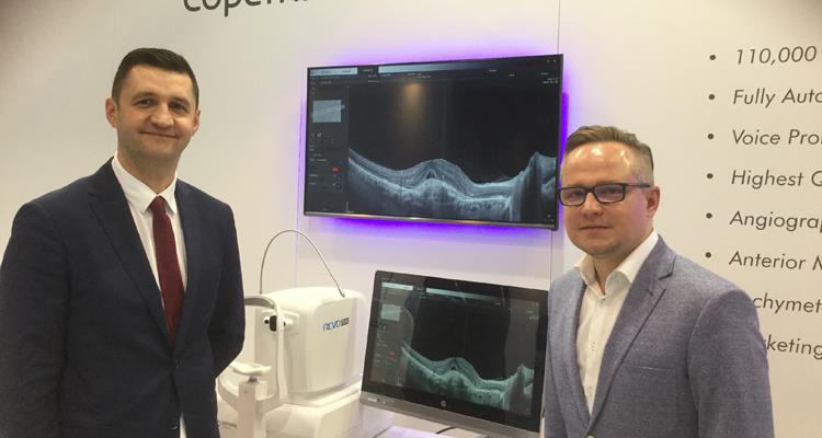
Figure 4
Topcon was displaying its Triton unit which is now becoming established as the go to swept source OCT and capable of high resolution imaging (figure 5) and also OCT angiography (figure 6). They also now have developed an intuitive software approach to disc analysis offering quantitative disc change assessment, again one we aim to review in more detail shortly.
Figure 5
Zeiss instruments had both the well-established Cirrus on display alongside the optometrist-friendly Primus, which is run from a slit-lamp table.
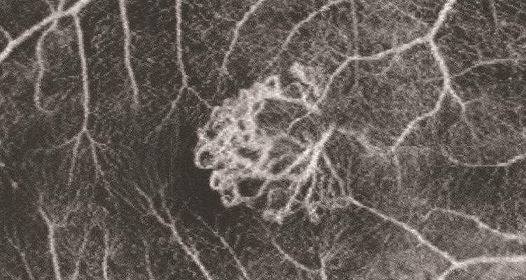
Figure 6
The Cirrus also has angiography options now and its data output into the Zeiss Forum software makes it easy to combine structural data with functional data such as from fields assessment, allowing a full assessment of each patient.
Haag-Streit UK was the first to show OCT angiography in the UK (figure 7) and the Optovue Angiovue was attracting much attention again at the show.
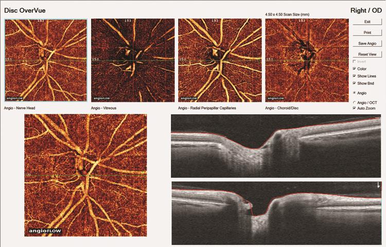
Figure 7
Digital imaging
But it was not all about OCT and there were interesting new imaging devices to be found throughout the exhibition. Carleton Optical distributes the ARC slit lamp camera unit which was also on display at Andrew Matheson’s stand (figure 8). The ARCcam HDSL IR adapter now offers higher resolution imaging and also includes a meibography option.
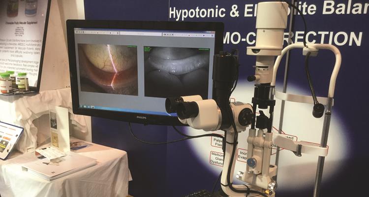
Figure 8
Optos had an impressive stand with its ultra-wide field scanning laser ophthalmoscopy devices, such as the Daytona and the California, much in demand. Behind the scenes there is a lot of research into these techniques as aiding the assessment of peripheral retinopathies and also in screening for amyloid plaques associated with some dementia states. Keep an eye out as this research becomes available later in the year.
The Kowa nonmyd WX3D (figure 9) was to be found at the Sense Medical stand. This camera seamlessly captures images for 3D display (something required in some screening and outreach clinic programmes) and is an instrument we will be highlighting in the coming weeks.
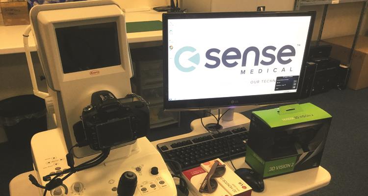
Figure 9
At the other end of the spectrum was the D-Eye camera phone attachment available from Grafton (figure 10). This simple to attach and operate camera adds to most modern smartphones and allows reasonable quality imaging for disc and central retina, and could be adapted for use out in the field where images might be transferred to a central clinic for analysis. A similar concept, but different design, is currently being launched by the charitable group Peek and, again, expect a more detailed review of this very soon.
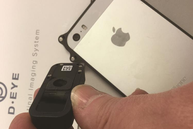
Figure 10
Still with the hand-held camera options, Mainline Optical was showing the latest incarnation of the Smartscope Pro (figure 11). This digital camera has been upgraded with much better resolution capture, the inclusion of adjustable fixation targets, and is now available with an optional slit-lamp adapter to ensure a steady capture.
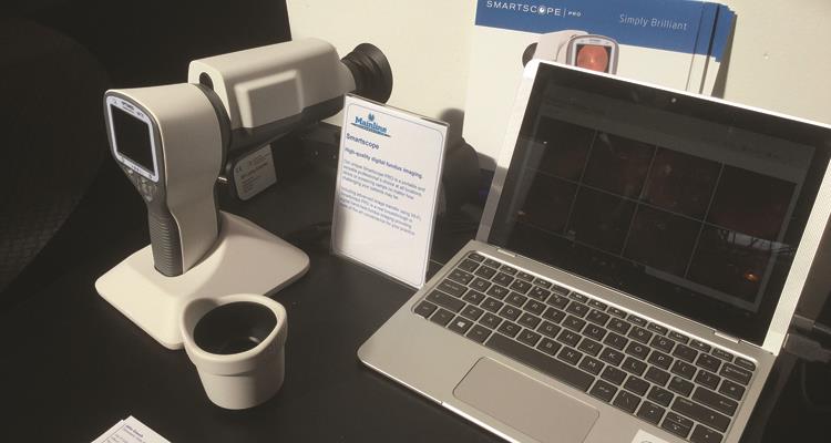
Figure 11
Dedicated slit-lamp imaging is increasingly popular and BIB was showing its newly launched slit lamp, the Righton LED Slit Lamp MW50D with integrated imaging system. The unit is capable of 50 times magnification allowing excellent image capture of anterior structures (figure 12).
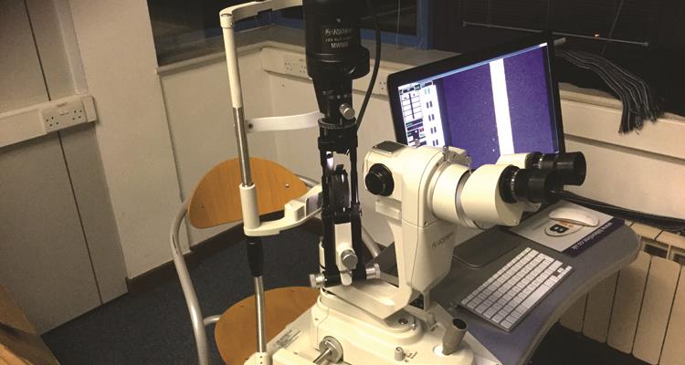
Figure 12
Another slit lamp imaging system of note is the Haag Streit FM300, a retinal image capture unit that can be used at the slit lamp and may be alternated with an anterior system to offer the clinician good all round image capture during the examination (figure 13). The unit sat well alongside the Eidon AF at the Haag stand, a true white light imaing system now capable of autofluorescence assessment.
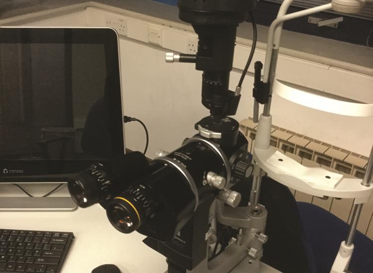
Figure 13
Image analysis
Expect to see an increased availability in software capable of automatically analysing captured images. One system that was being previewed at the Birmingham Optical Group stand is the new Advanced Ophthalmic Systems (AOS) system. This software allows any captured images to be analysed in a variety of ways, such as the counting and grading of corneal stain, assessment of vascularisation and hyperaemia and the analysis of retinopathic changes (figures 14 a to d). I hope to be trialling this shortly but initial impressions are that this may well make image analysis more robust and objective and remove inter-practitioner subjective variation.
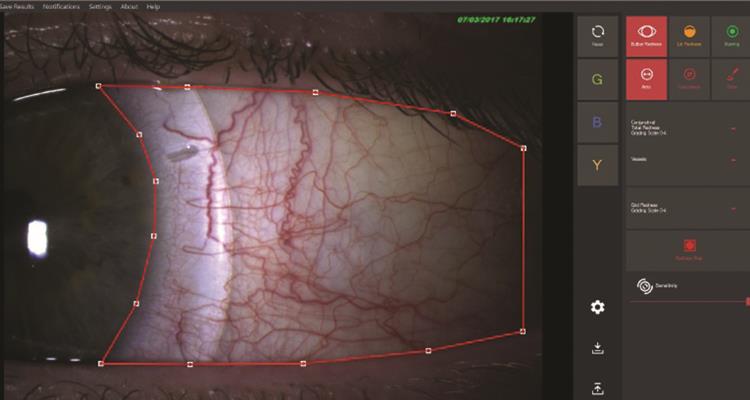
Figure 14a
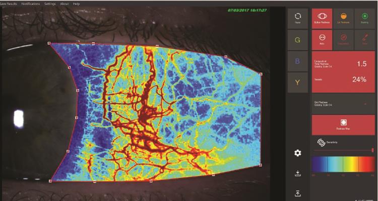
Figure 14b
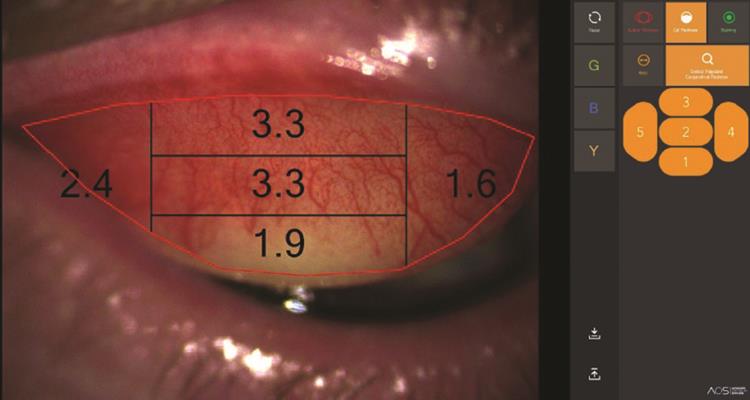
Figure 14c
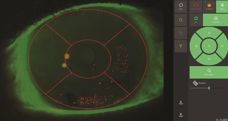
Figure 14d
New technologies
There were two systems on display that represent new ways of assessing the eye and both of which may well become part of future eye assessment. Eye tracking technology has come on leaps and bounds in recent years and the Clinical Eye Tracker developed by Thomson Software Solutions was attracting much interest (figure 15).
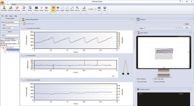
Figure 15
As inventor Professor David Thomson told me: ‘The new system, which was shortlisted for the AOP Product of the Year Award, allows clinicians to assess binocular vision and eye movements while the patient carries out normal visual tasks such as reading, tracking, searching and so on. As one visitor to the stand commented, “this brings binocular vision assessment into the 21st century”.’
Equally interesting is the new electrophysiology system available from DiopSys. I first came across this in the US and was impressed by the ease with which objective data about the functioning of the retina and visual pathway may be acquired via one simple electrode attachment (figure 16). When you consider the variability inherent in standard fields assessment due to patient subjective responses, this system surely must be of interest to optometry. Expect further discussion of this system later in the year.
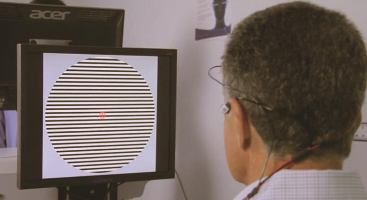
Figure 16
Dispensing is to benefit from new technology too. Hoya was showcasing its new Yuniku system (figure 17). This digital dispensing unit quickly and accurately measures the patient, and then emails the data to the laboratory where the spectacles can then be 3D printed, ready for supply back to the practice. This is likely to be a pioneer for much future dispensing in my humble view.
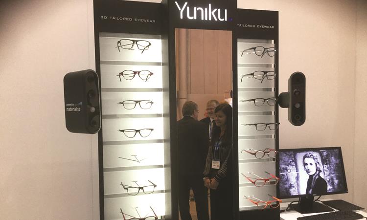
Figure 17
Others
Carleton Optical showed the latest incarnation of its excellent Plusoptix paediatric screening unit, the S12 (figure 18). This binocular autorefractor has proved itself in paediatric screening programmes and the new system offers updated software aiding the referral and reporting of findings. There was also much interest surrounded the E-Eye unit from Grafton which we recently reported on (Optician 17.03.17). I hope to run a case study on this unit shortly.
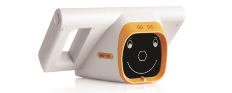
Figure 18
Visual fields testing was not forgotten and Birmingham Optical Group (BOG) was showing the latest fields screener from Oculus, the Smart Field, with its impressively small footprint which should make it a valuable investment for those with restricted space in their practice (figure 19).
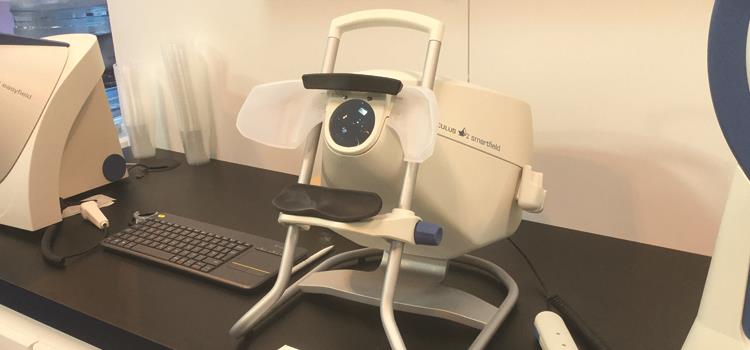
Figure 19
BOG was also launching a new electronic acuity system with an impressive array of simulated views (figures 20 a and b).
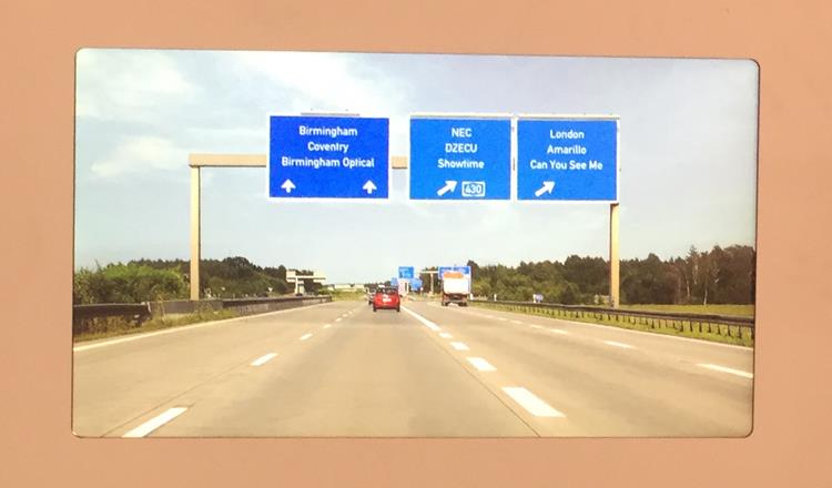
Figure 20a

Figure 20b
Keeler is focused on its centenary at present but talk at the stand suggested a major development in their tonometer range coming very soon.
Thomson Software Solutions was showcasing a new version of its ReadEZ screening software. The software measures symptoms and reading speed against a white background then uses an algorithm developed by Professor Thomson to determine the optimum colour for overlays, clip-ons, prescription lenses and computer screens. The latest version adds a second level of saturations for the coloured lenses and the ability to prescribe bespoke tints.
Altacor, a company perhaps best known for their AREDS-friendly chocolate bar, is now venturing into the punctum plug market with a new dissolvable plug that fits inside the punctum and looks a promising addition to the available range.
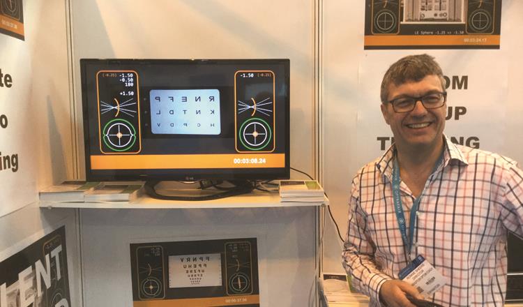
Figure 21
And finally promoting the view that we all learn from our own mistake, ‘LenShuffler’ (figure 21) was encouraging everyone to check out his uploaded youtube videos which follow different practitioner’s assessments. He is happy to film readers and offer personalised feedback and coaching.