
I do not think I am alone in thinking that how binocular vision was taught at undergraduate level did not always make this subject either clear or inviting. My understanding of the topic really only developed once in the real world and, particularly, after becoming more confident when dealing with young children, a patient group I was initially terrified of on first qualifying.
The first in our interactive exercises in binocular vision was designed to be an introduction to some more challenging scenarios we will publish in future months. It was aimed at encouraging an understanding of the main assessment techniques and understanding the possible causes of unilateral vision reduction.
Case Scenario
A mother brings her son to you for advice about his vision. He has just turned five years old, and has been at school for the best part of three terms. She is worried because the school have suggested he has an eye check because they feel he may have poor vision in one eye. There is no report of any ‘turn in the eye’ and initial gross observation confirms this.
The boy is healthy, had a normal full-term birth and today seems both relaxed and unduly concerned about the attention afforded him.
Furthermore, there is no family history of any ocular or optical concerns.
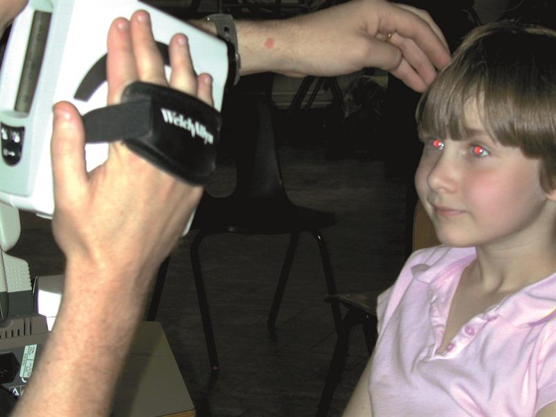
Using the most appropriate vision test, the following is found:
Right 0.3 logMAR Left 0.0 logMAR
Consider the following:
• What further tests would be needed to ascertain the likely cause of this asymmetry, and how might you explain them to the concerned mother?
• There are a number of possible refractive or binocular anomalies that might cause this vision difference. What are they, and how could you explain each in a way the mother would understand?
Further Tests
Perhaps because of the point being made that initial gross observation confirmed no turn, many respondents did not mention the cover test in the assessment, something I think you will agree, would be essential in the initial assessment, after accurate visions and before refraction (most agreed pre and post cyclo). About half of respondents mentioned stereopsis assessment (something we will take a closer look at in the next of our CET series), and most included tests specific to finding out any possible pathology or binocular anomaly (such as a 4Δbase out test for a microtropia). Here are a few examples of response.
‘Assuming that this is the unaided vision, the first step would be to refract to see if a prescription in the right eye would improve the vision. If not, keratometry, topography and slit lamp examination would reveal any significant corneal irregularities. Then ophthalmoscopy would investigate for any lens opacities and retinal abnormalities. A fundus photo and OCT might be useful.’
‘Full cycloplegic examination, alternating cover test and motility and stereopsis assessments.’
‘Do a cycloplegic and, prior to that, check distance and near OMB [presumably this means a cover test] and motility, pupils and near acuity.’
‘The further tests needed would be a stereopsis test such as a Titmus fly. A cover test and cycloplegic refraction would be appropriate. Finally, fundoscopy using an ophthalmoscope or indirect ophthalmoscopy.’
Explanation
There were some excellent contributions regarding explaining tests to the mother, something that would prove essential if compliance with any management was required in the future. Here are some examples.
‘I would explain to the mother that his vision is below the age matched normal in his RE. This could be due to an uncorrected refractive error or due to amblyopia. I would explain to the concerned mother that further tests are needed for a diagnosis.’
‘Each step would be explained to the concerned mother in simple terms, as would the likely chance of improving the vision. It would be easy to explain to the mother if a low power prescription (-1.00 for example) was all that was necessary to correct the right eye. Obviously, if there was some pathology impairing the vision, that would need a more subtle approach. Then if some orthoptic treatment or prism correction was necessary, we would need to explain in more detail the reasons and that this is more likely to be an ongoing process.’
‘Subjective refraction is simply asking the child to look at a letter chart with each eye individually and asking him what letters he is able to read. Then putting different strengths of lenses over his eyes to see if the vision improves. Retinoscopy would involve shining a light into his eyes. From this, the optometrist can get an idea of the prescription and also examine the back of the eye to see if the lens is cloudy or if there is anything usual about the back of the eye. Cycloplegic refraction would mean putting drops into her son’s eyes which will sting a little, but would allow a much better examination of the back of the eye and may also help to decide if a prescription is necessary. The 4Δbase out test would just involve a lens being put in front of her sons eye to see whether the second eye would react to it [to confirm a microstrabismus].’
‘Demonstrate a +2.00DS sphere in front of mum’s eye for amblyopia, then a 2.00DC cylinder obliquely for meridional amblyopia. Then show her a small prism so the vision just splits to demonstrate a small strabismus.’
Possible Causes
The vision in the right eye is reduced by three lines on the logMAR chart (to 6/9 Snellen equivalent). If this is purely refractive, a pinhole might be useful to check for improvement. And the cover test would rule out any strabismic element. Assuming orthophoria, the reduction might be:
• Uncorrected myopia (around -0.75DS)
• Uncorrected cylinder (1.50DC or so)
• +0.75DS fogging in the binocular state with established amblyopia (so little improvement in the monocular state if accommodating). Cycloplegia would be important to establish full correction influence here
If the reduction is strabismic, the low level reduction would imply a small strabismus and it is likely that there might be some sensory adaptation such as anomalous retinal correspondence. Thus, checking for a microstrabismus would seem appropriate.
Typical of your summaries was: ‘Possible causes may be due anisometropia. A large [sic] uncorrected refractive error in the RE may be causing the reduced vision. Another cause may be amblyopia due to a squint (microtropia if difficult to detect).’ Another, ‘Possible micro strabismus, meridional amblyopia, anisometropia.’ And let us not forget the, albeit not too likely, possibility of some disease process (congenital or developmental) reducing vision in the right eye.