Cardiff School of Optometry and Vision Sciences hosted a festschrift last month to celebrate and honour the achievements of a highly respected member of the research community. Cameron Hudson reports
Professor Neville Drasdo's 'festschrift' (from the German 'fest' - celebration and 'schrift' - writing) attracted over 80 delegates and comprised a series of CET accredited lectures presented by some of Professor Drasdo's former colleagues, PhD students and co-authors.
Throughout his academic career, spanning more than 50 years, Professor Drasdo has made enormous and invaluable contributions to vision science research. Through his exceptional academic ability and great character he has become a highly respected and much loved member of the research community.
Professor Drasdo began his academic career in 1961, when he was appointed as lecturer in ophthalmic optics in the Physics Department at the Birmingham College of Advanced Technology, which was later to become Aston University. Through his enthusiasm to promote and extend vision science knowledge, he set up the Master's course in Methods of Ophthalmic Investigation, which eventually achieved the distinction of MRC recognition.
His research interests at that time were on a wide range of subjects, varying from corneal physiology to infant visual development and his collaborations with neurophysiology were instrumental in the formation of the Aston University, Department of Vision Sciences.
During his time as clinical tutor and PhD supervisor, Professor Drasdo successfully turned out 42 MSc and 11 PhD graduates, many of whom have since become successful researchers and academics.
In 1993, Professor Drasdo received the academic degree of Doctor of Science and shortly after almost simultaneously became Dean of Life Sciences at Aston University as well as participating in the Research Assessment exercise.
During more recent years Professor Drasdo moved to the Cardiff School of Optometry and Vision Sciences where his research expertise,
particularly in the field of electrophysiology, has been an invaluable asset to the department.
Many of the speakers during the event took the opportunity to thank Professor Drasdo for his inspiration, enthusiasm and contributions to their own careers as well as to vision science research.
OPTICS AND PSYCHOPHYSICS
In the first lecture of the day, titled 'Refraction and retinal image quality in the peripheral field', Professor Neil Charman, (Faculty of Life Sciences, University of Manchester) described how the optical performance of the eye governs visual performance across the visual field. This relationship was pointed out by Professor Drasdo many years earlier. However, with the use of sophisticated aberrometers and autorefractors, Charman's group were able to determine image quality and refractive error to field angles of 30 degrees or more.
Professor Charman briefly reviewed how optical performance in the peripheral field may be a governing factor for axial refractive error.
Professor David Whitaker (Department of Optometry, University of Bradford) presented 'Gradients in peripheral vision: diversity within parallel chromatic pathways'. Professor Whitaker presented a study of the chromatic sensitivity in central and peripheral vision for the parallel visual pathways, namely the blue-yellow (S/L+M) and red-green (L/M) as well as the achromatic pathway (L+M). Using a method of spatial scaling first described by Professor Drasdo in 1991, Professor Whitaker's group was able to determine contrast sensitivity functions for stimuli designed to isolate each of the above pathways.
Interestingly, the results indicate that while both chromatic parallel pathways obeyed the concept of spatial scaling, the two chromatic scaling factors differed by almost an order of magnitude. The findings of this study were deemed to have critical bearing upon the physiological debate regarding the specificity or randomness of cone connectivity within chromatic mechanism.
Dr Ian Murray (Faculty of Life Sciences, University of Manchester) gave the third lecture 'Discounting the illuminant: how stable is our colour vision?' Dr Murray described one of his studies that employed successive asymmetric colour matching tasks to investigate the changes in colour appearance of simulated colour samples when colour shifts were induced with two Planckian illuminants (A and S). The results of his study suggested that human colour vision was more stable when viewing conditions were unrestricted and longer periods of adaptation were used (up to 60 seconds).
The final lecture of the first session titled 'Ricco's area for 'on' and 'off'
S-cone stimuli' was given by Professor Roger Anderson (School of Biomedical Sciences, University of Ulster, Coleraine). He described how his group was able to measure the detection threshold of the retinal pathways transmitting S-cone 'on' and 'off' signals. The detection thresholds of stimuli that varied in size and retinal location were then used to determine the subtype of ganglion cells responsible for their detection.
PSYCHOLOGICAL AND APPLIED INVESTIGATIONS
The first lecture of the second session was given by Professor Robert Hess (Department of Ophthalmology, McGill University, Montreal, Canada). He gave an interesting talk entitled 'The site and nature of the cortical deficit in human amblyopia' and described how clinical opinions had shifted from amblyopia being considered simply as a deficit to contrast sensitivity. He demonstrated that the origins of amblyopic deficit could be localised to extrastriate areas of the cortex, through investigation of the second-order processing mechanisms in cases of acquired cortical pathology and amblyopia. His research, using both human psychophysics and brain imaging, indicates that amblyopia largely affects the integration of visual information for second order, in particular motion and stereo processing with compelling evidence for extrastriate rather than striate cortical deficit, as previously thought.
The second, more light-hearted presentation of the session came from Professor Richard Adabi (Faculty of Life Sciences, University of Manchester). Professor Adabi gave a humorous but informed talk on the perception of motion and how the brain differentiates retinal image movement produced by an object or scene, from image motion secondary to eye movements. He described one of the techniques he had used to artificially interrupt the normal sensory-motor control loop, in patients with nystagmus. Put simply, the coordination of stimulus movement with eye movement in patients with nystagmus, led to the perception of motion. The results of these findings show that extra-retinal signals are primarily responsible for spatial constancy during visual search eye movements.
Professor Gordon Heron (Department of Vision Sciences, Glasgow Caledonian University) presented a lecture entitled 'The Pulfrich effect in practice'. Professor Heron gave several examples of how the Pulfrich effect may be responsible for disturbing visual symptoms arising from errors in the mislocation of objects in space. Such abnormality, he noted, could often be seen in patients with uniocular latency delays as in multiple sclerosis or mid facial trauma.
The Pulfrich effect can be demonstrated in the normal eye by placing a filter over one eye while viewing an obliquely swinging pendulum whereby apparent depth becomes evident. It was pointed out that sufferers of this effect may encounter difficulties pouring liquids into vessels, putting keys in locks etc but potentially more harmful difficulties arise when travelling in cars. In a similar way that the effect can be stimulated in the normal eye, Professor Heron's group had been successful in removing the effect in two thirds of symptomatic subjects through the provision of a suitable ophthalmic tint before the least affected eye. The results of this study may have interesting implications for treatment of such symptoms in clinical practice.
Speaking on the 'Influence of age on vision' Professor David Elliot (Department of Optometry, University of Bradford) addressed some controversies in this area. He highlighted that the greatest uncertainty was the principal cause of visual reduction with age. He argued that despite the common perception regarding optical factors such as increased light scatter, increased lens absorption and pupillary miosis, the principal causes may instead be due to neural factors. Professor Elliot's group was able to demonstrate that optical factors did not contribute to an overall increase in ocular aberration with age since the 'beneficial' effects of age-related pupillary miosis were sufficient to eliminate the effects of light scatter and lens absorption.
Dr Colin Fowler (School of Life and Health Sciences, Aston University) gave a presentation titled 'Eyes and aspherics'. As a former PhD student of Professor Drasdo in the early 1970s, Dr Fowler first explained that through his and Professor Drasdo's initial interest in the investigation of peripheral aberrations on visual perception they were able to create a model eye using a simple aspheric cornea. He then described how aspheric surface design had evolved with the advancement of computer technology. He highlighted the relevance of such technology when applied to progressive spectacle lenses and refractive surgery.
The final lecture of the session was given by Professor Bernard Gilmartin (School of Life and Health Sciences, Aston University) who spoke on 'Myopia: a new dimension'. Professor Gilmartin addressed the epidemiological prevalence of myopia and the conflict between genetic and environmental factors for myopia development. Using MRI-based imaging techniques Professor Gilmartin has been able to model the human eye in three dimensions and demonstrated how such technology may address such intriguing questions concerning the effects of central and peripheral stretch on ocular aberrations, the relationship between receptor orientation and image quality and the clinical assessment of ocular risk in high myopia.
CLINICAL ISSUES
Following a buffet lunch the third session was aimed at addressing clinical issues. Professor Bruce Evans (Institute of Optometry), also one of Professor Drasdo's former PhD students, gave the first of the afternoon presentations titled 'Linking the visual deficits in dyslexia: an evolving hypothesis'. Professor Evans first addressed some of the best established visual correlates of dyslexia which included binocular instability, Meares-Irlen syndrome, pattern glare and a deficit of the magno-cellular sub-system.
Professor Evans pointed out the recent interest towards a visual attention deficit as a possible correlate for dyslexia and how visual attention may in fact underlie many of the more popular correlates. Professor Evans concluded by complementing Professor Drasdo for his tremendous foresight in highlighting a deficit in visual attention as a causal factor in dyslexia as reported in his first contribution to the subject over 33 years ago.
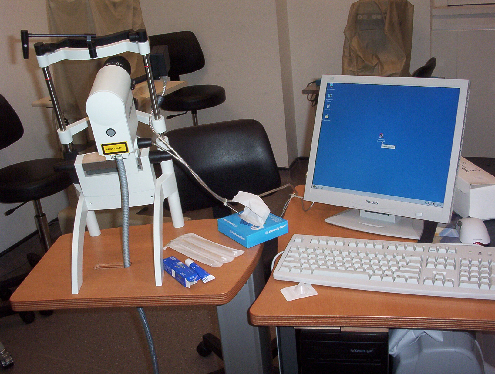
Professor Rachel North (Cardiff School of Optometry and Vision Sciences) spoke on 'Digital imaging and electrophysiology in glaucoma and ocular hypertension'. Professor North presented the results of a study in which the photopic negative response (PhNR), pattern electroretinogram (PERG) and the S-cone ERG were correlated morphological optic nerve head measurements made using optical coherence tomography (OCT) and the Heidelberg retina tomograph (HRT) in glaucomatous, ocular hypertensive and normal individuals. The study suggested that functional changes precede detectable structural alterations to optic nerve head morphology and that the earliest changes in ganglion cell death may in fact be reversible.

The HRT II was used by Professor Rachel North to detect structural changes in a study of glaucoma and ocular hypertension
The third, and eagerly anticipated, lecture of the afternoon was given by Professor Drasdo on 'A psychophysically based model of receptive field
density: a required necessity for assessment of neural damage in glaucoma and ocular hypertension'. Professor Drasdo described a technique by which the receptive field density could be calculated by a complex calculation based upon the E2 parameter for minimum angle of resolution of a grating. Professor Drasdo stumbled across this calculation in the mid 1970s. However, it was not until more than a decade later that a similar study was able to generate data which confirmed his early estimates.
Professor David Henson (Manchester University School of Medicine) addressed some important issues relating the clinical utility of current research in glaucoma in a lecture entitled 'Glaucoma; realigning research priorities'. Professor Henson reviewed epidemiological studies to determine whether glaucoma research to date had in fact been of any benefit to sufferers. He highlighted that the three principal causes of blindness from the disease were aggressive disease, treatment compliance problems or advanced disease at the time of presentation. Since the incidence of glaucoma had remained stable over the past two decades, he suggested that less emphasis should be put on early detection and attention redirected towards the principal factors for blindness from the disease.
The final speaker of the session Professor Geoff Arden (Department of Optometry and Vision Science, City University) spoke on 'Relative anoxia in the apparently normal diabetic eye: a possible important factor in the production of retinopathy'. He described how administration of oxygen to patients with diabetes helped to restore tritan visual deficit even in patients with normal fundi and normal acuity. During darkness the rod cell dark current is very demanding on retinal oxygen levels leading to relative anoxia under such conditions. He described novel experiments using diabetic patients with retinitis pigmentosa and showed that in these patients retinopathy did not develop to the abolition of the dark current. He concluded by suggesting several novel ways in which the oxygen demand of the retina may be reduced and/or controlled.
CORTICAL IMAGING AND VISUAL ELECTROPHYSIOLOGY
The final session of the day began with a lecture from Dr Paul Furlong (School of Life and Health Sciences, Aston University) on 'Brain mapping'.
He acknowledged the seminal work of professors Drasdo and Harding during the 1980s and how their influence inspired a generation of researchers to push the frontiers of such techniques. Dr Furlong described how modern spatial filter magnetoencephalography (MEG) techniques, while still in their infancy, could be used as a tool for cognitive evaluation in providing estimations of the location of neural activity in the brain with much promise of a clinical application in aiding neuronavigational techniques during brain surgery.
The second lecture of the session was presented by Professor Stephen Anderson (School of Life and Health Sciences, Aston University) entitled 'The spatial distribution and temporal dynamics of brain areas activated during the perception of objects'. Using MEG and magnetic resonance imaging (MRI), Professor Anderson explained how his research group was able to source in the brain those regions governing the motor affordance of a grasping action in response to different patterns of visual perceptual input.
Professor Daphne McCulloch (Department of Vision Sciences, Glasgow Caledonian University) presented 'Maturation of VEPs in human infants: is there really a gender difference?'. Professor McCulloch highlighted the clinical utility of the VEP in the assessment of infantile vision through its objective and relatively non-invasive nature. Professor McCulloch's group had measured distinctly earlier VEP maturation in female infants.
These differences in the VEP corroborate well with other known and well documented gender differences in the neurological and cognitive development of infants.
The final lecture of the day was presented by Dr Dorothy Neville (Great Ormond Street Hospital for Children, London). As one of Professor Drasdo's former PhD students, Dr Neville described some of her earliest experiences in visual electrophysiology and how she has come to employ similar techniques in various studies attempting to understand why visual development goes wrong.
After the lecture series delegates were invited to attend an informal celebratory dinner which gave many of Professor Drasdo's former colleagues and acquaintances the opportunity to enjoy, honour and celebrate his notable career and research achievements.
The Cardiff School of Optometry and Vision Sciences was grateful to Carl Zeiss UK, Essilor, Keeler, Topcon Great Britain and Cambridge Research Systems for sponsoring the event.
