Wavefront Lasek
Case history (DOB: 24/11/79)
The Lasek (laser assisted epithelial keratomileusis) technique is a surface based treatment where a thin flap of epithelium is created (~60<03BC>m). The flap in Lasik is much thicker (~160<03BC>m) and the technique more invasive. Lasek is therefore often used in thinner corneas or where it may be difficult to use a microkeratome.
The patient was an RGP then soft contact lens wearer for 7 years but had ceased contact lens wear 18/12 ago and now only uses them for social wear. Motivation for treatment was due to poor vision quality with lenses and glasses.
There were no other significant ocular or general health complications.
Preoperative refraction
| UCVA | Sph | Cyl | Axis | BCVA | |
| R | 6/120 | -8.25 | -1.00 | 175 | 6/5 |
| L | 6/120 | -8.00 | -1.25 | 170 | 6/5 |
Bin: UCVA 6/120 - Bin: BCVA 6/5
Clinical information
Scotopic pupil size: R 7.6 mm L 7.6 mm, measured with the Neuroptics pupilometer. This allows for the measurement of dynamic pupil size
Pachymetry: (ultrasound) R 485µm L 487µm
Orbscan keratometry readings:
R 46.8D x 98 Max L 47.1D x 74 Max
R 45.8D x 8 Min L 45.6D x 164 Min
The Orbscan provides information on elevation of the anterior and posterior cornea in addition to axial keratometric maps. This provides detailed information on whether it may be safe to perform laser refractive surgery.
Biomicroscopic examination: Anterior segment clear both eyes.
Wavefront aberrometry (Figures 1and 2): The bottom right hand images on these maps represent the point spread function (PSF). This shows the distortion of a 'pinpoint' of light as it hits the foveola due to higher order aberrations, and is one of a number of selection criteria used for wavefront.
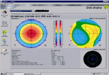 |
| Figure 1 |
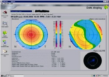 |
| Figure 2 |
Treatment plan
Bilateral wavefront Lasek was chosen as there was not sufficient tissue for Lasik.
Special points emphasised during the consent procedure were potential undercorrection, delayed healing, pain and haze and the need for critical distance glasses postop.
| Sph | Cyl | Axis | Optic Zone | |
| R | -8.25 | -8.25 | 175 | 7 |
| L | -8.00 | -1.25 | 170 | 6.9 |
Routine surgery was carried out, with no complications both eyes. Bandage silicone hydrogel lenses inserted in both eyes to protect the delicate epithelial flap for the first four days.
Follow up
ONE DAY: No discomfort or pain Vision: R 6/24 L 6/24 BIN 6/18 with plano bandage CLs in situ.
Biomicroscopic examination: CLs in situ, moving well. It is important to ensure that the bandage CLs move in order to facilitate easy removal. Patients are asked to use artificial tears to aid this
FOUR DAYS: Both eyes felt gritty. Bandage lenses removed, after confirming movement was good (no binding)
Vision: R 6/24 L 6/24 BIN 6/18 NB it is normal in Lasek for the vision to be low when the contact lens is first removed, as the epithelium has not yet settled Biomicroscopic examination: R 10 per cent epithelial defect, L 40 per cent epithelial defect centrally. Action: The following medication was prescribed, to aid healing and dry eye. Maxidex and Exocin qds, Visilube 6/day, Snotears 6/day, Volterol bds
ONE WEEK: R doing well, L still gritty Vision: R 6/7.5 L 6/60 Biomicroscopic examination: R cornea smooth and clear with a trace of superficial punctate keratopathy (SPK). L SPK and central epithelial defect, affecting vision Action: Gel tears 6/day, L Lacrilube bds
TWO WEEKS: Both eyes doing much better, though L still more uncomfortable than R, and some dryness in both
Refraction:
| UCVA | Sph | Cyl | Axis | BCVA | |
| R | 6/7.5 | +1.00 | -0.50 | 12 | 6/6 |
| L | 6/15+ | +2.00 | -0.50 | 76 | 6/9+ |
Biomicroscopic examination: R smooth and clear, L moderate SPK but no epithelial defect present.
Trace haze LE Action: Gel tears 6/day Lacrilube bds BE.
EIGHT WEEKS: Minimal dryness and patient was very pleased with results.
Refraction:
| UCVA | Sph | Cyl | Axis | BCVA | |
| R | 6/5 | +0.50 | -0.50 | 12 | 6/5 |
| L | 6/7.5+ | +0.75 | -0.50 | 80 | 6/5 |
Biomicroscopic examination: Clear R, L trace haze as before.
Conclusion
This was an interesting case, demonstrating that Lasek with a high Rx can occasionally give delayed healing (as with the left eye). However in time both eyes settled well, and Wavefront technology made this treatment safer and more predictable than using a Planoscan technique.
- Emma Firmager is an optometrist and professional services manager for Advance VisionCare. The surgery was crried out by Mr CT Pillai.