John Bourton and Deacon Harle describe an interesting presentation at the Institute of Optometry Clinic
An asymptomatic recently diagnosed (2004) type II diabetic, 54-year-old female of Asian ethnicity attended for a routine annual eye examination.
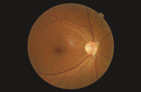 |
| Figure 1. A Bergmeister's papilla |
A Bergmeister's papilla is a congenital anomaly histochemically defined as part of the common hyaloid system.1
During gestation a proliferation of glial cells from the optic disc creates a peri-vascular sheath around the hyaloid artery.2,3 The mechanism responsible for the atrophy of the hyaloid artery before birth can fail, allowing remnants to remain. A persistence of the central hyaloid artery in whole or part is one of the commonest developmental anomalies occurring in the human eye.4 Posterior remnants may occur at the disc in some 95 per cent of premature infants and 3 per cent of full-term infants. The form and extent of the persistence can show a wide range of variation and in this case has a surprising and unusual triangular appearance.
Most findings of Bergmeister papillae are incidental and have no visual significance, but occasionally the condition can be associated with retinochoroidal coloboma5,6 and general developmental disorders such as Potter's syndrome7 and Edward's syndrome.8 Differential diagnosis necessarily should exclude tumour, myelinated nerve fibres and posterior vitreous detachment.
References
1 Zhu M, Provis JM, Penfold PL. The human hyaloid system: cellular phenotypes and inter-relationships. Exp Eye Res, 1999; 68(5):553-63.
2 Rhodes RH. Development of the human optic disc: light microscopy. Am J Anat, 1978; 153(4):601-15.
3 Hamming NA, Apple DJ, Gieser DK, Vygantas CM. Ultrastructure of the hyaloid vasculature in primates. Invest Ophthalmol Vis Sci ,1977; 16(5):408-15.
4 System of ophthalmology Vol III Part 2, Congenital Deformities. Edited Sir Stewart Duke-Elder Page 765 29-2.
5 Giuffre G. Remants of Bergmeister's papilla and retinochoroidal colobomas. Ann Ophthalmol, 1987; 19(8):16-8.
6 Connolly WE, Polomeno RC. Optic disc colobomas. Can J Ophthalmol, 1983; 18(6): 299-301.
7 Brownstein S, Kirkham TH, Kalousek DK. Bilateral renal agenesis with multiple congenital ocular anomalies. Am J Ophthalmol, 1976; 82(5):770-4.
8 Pe'er J, Braun JT. Ocular pathology in trisomy 18 (Edwards' syndrome), Ophthalmologica, 1986; 192(3):176-8.