 |
This article has been adapted from Routine Eye Examination by Bill Harvey and Andy Franklin, part of the new Eye Essentials series. ORDER YOUR COPY |
To mark the launch of a major new series of textbooks, Optician begins a new regular CET series with a short article by Bill Harvey and Andrew Franklin outlining some key points about refraction.
CET module C1631. This module has now closed.
Objective refraction
Objective refraction includes retinoscopy and the use of autorefractors, which are appearing in optometric practice in increasing numbers. In general, objective methods are not required to give us a final prescription.
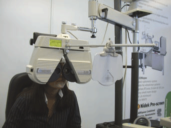
They merely need to get us to a point from which subjective methods can take us to the end point accurately and quickly. Increasingly, phoropter heads are used instead of trial frames.
 |
|
Phoropter heads are a useful aid |
Modern automated lens carriages allow fast lens insertion, greater accuracy in axis location, more rapid comparison presentation of lenses, use of variable prism, and a variety of further options depending on the model, such as immediate comparison of new refractive findings with previous results.
They are not advisable for a few situations, notably low vision assessment and over-refraction of multifocal contact lenses, where the reduced light levels may affect visual performance or pupil size respectively.
RETINOSCOPY
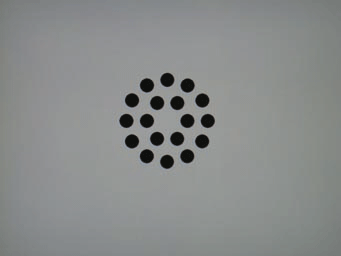
Target
Ideally, we want a target that will promote accurate and steady fixation, but no stimulus to accommodation.
Various targets are used and they probably make little difference to the end result, but there is some evidence that the rings on the green block of the duochrome might be the version that produces least accommodation.
In the absence of any contradictory evidence the rings on the green would be the recommended fixation target for retinoscopy.
Fogging
During retinoscopy, it is the fixating eye that controls accommodation, so it must be fogged to ensure that accommodation is relaxed. However, if you overdo it, you can induce accommodation, so the fogging should be less than 2.00D.
Initially, both eyes should be corrected with what you think is likely to be the full plus correction, based on the patient's existing correction (if available), VA and symptoms and history, plus the working distance allowance.
Check with the retinoscope that you have an 'against' movement in either eye. From time to time during retinoscopy, pass the retinoscope beam across the fixating eye to make sure that it is still fogged. This is particularly important with hyperopic patients.
Initial lens
If you have the patient's last spectacle prescription, this is a good starting point. If the patient has lost their spectacles or no previous prescription is available, consider the unaided vision and far point.
A little thought at this stage can save a lot of time and effort. There is nothing to stop you checking the VA when you have neutralised the more positive meridian, to get an idea of the cylinder power required.
Distance unaided vision is related to refractive error in myopes and manifest hyperopes (Table 1).
| Table 1. Relating the vision to the mean sphere | ||||||||||||||||
|
It is also the case in purely astigmatic refractive errors - or with the best vision sphere in place (Table 2).
| Table 2. Relating the vision to the uncorrected cylinder | ||||||||||||
|
It is important to remember that these are only average values. Patients with small pupils (usually presbyopes) will experience less blur per dioptre, and those with large pupils more.
For myopes, the far point at which small print can be seen clearly varies inversely with refractive error (Table 3).
| Table 3. The far point for uncorrected myopia | ||||||||||||||
|
The working lens should be incorporated into the correcting sphere. The use of a separate working lens introduces an extra set of reflections and it uses up one of the trial frame spaces you may need for the patient's prescription.
For the ideal starting point, we would want the patient slightly fogged (over-plussed) for distance to discourage accommodation, but rather less than 2.00D. The easiest type of reflex to interpret is a quick 'with' movement which should occur if the patient is slightly under-plussed for your working distance.
They will still be somewhat fogged for the distance that the patient is fixating as your retinoscope and the patient are separated by 1.50D.
Plus or minus cylinder?
Plus cylinders do tend to give a clearer, more easily neutralised streak and are favoured by some practitioners who rely on retinoscopy to provide a final prescription (eg working with special needs patients).
However, using minus cylinders does ensure that accommodation is more easily controlled and most automated refractor heads do not have a plus cylinder option. Minus cylinders are more commonly used in routine refraction.
Streak or spot?
Either type of retinoscope will do the job, in the hands of someone familiar with it. Streak retinoscopes are currently fashionable and they make axis determination easier where there are high cylinders, but spot retinoscopes probably make it easier to do lower levels of astigmatism.
Some retinoscopes now come with a choice of bulb, so practitioners can experiment for themselves.
Autorefractors
Most studies suggest that autorefractors are quick, simple, repeatable and accurate (with some qualification). With cycloplegia or good accommodative control the results are very accurate. Indeed the spherical aberration introduced by the dilation of a cycloplegic makes the method preferable to retinoscopy in many cases.
Its ease of use makes it suitable to be carried out by ancillary staff, so reducing the burden of the optometrist. The machines may directly link to an automated phoropter head, again making the routine refraction more fluid.
It is useful to remember that even the most accurate objective measurement may not be that preferred by the patient, so a subjective approach is always preferable to ensure a tolerable refractive error, even though this is sometimes modified away from the actual refractive error present.
Subjective refraction
With retinoscopy completed we can proceed to the subjective stage of refraction. In an ideal world, what you now have in the trial frame is the patient's full spectacle correction plus your working lens.
The accurate correction of the astigmatic error requires that the 'circle of least confusion' (CLC) is placed on the retina and kept there while the cross-cylinder is in use. If this is not achieved, both the axis and power found will be wrong.
The range of spherical powers which allow maximum acuity can be several dioptres in patients with small pupils and low acuities, though it is usually smaller.
There may be a unique value for the correction which gives maximum contrast, though where accommodation is active this may also be a range. In this latter case the most positive lens that gives maximum contrast is usually the optimum correction.
The recommended routine includes a rather elaborate sequence of checks on the sphere power before, during and after the cross-cylinder is used. This is to ensure that:
Binocular vs monocular refraction
Autorefractors are appearing in optometric practice in increasing numbers
Binocular refraction has no disadvantages when compared to monocular refraction, though there are patients who cannot be refracted binocularly.
Advantages of binocular refraction
Limitations
Binocular refraction should not be used where acuities are markedly unequal, or one eye is strongly 'dominant'. If you do start to apply it to an unsuitable patient, they will usually tell you fairly quickly ('should I be seeing double?').
In such cases, occlude the better eye, refract the worst eye. Then refract the better eye with the worst eye fogged, if necessary, or even unfogged if the acuity is considerably worse than the eye you are going to test. If the worst eye is 6/9 or less, it may be unnecessary to fog it.
If the difference is less it is often possible to refract binocularly, but the final decision has to be taken on an individual case basis.
To place the CLC near the retina
Initially, we must determine the 'best vision sphere' (BVS), which can be defined as the most plus or least minus lens with which the patient can enjoy maximum visual acuity. In order that we do not under-plus, a 'fogging' technique is used. For each eye:
To refine the best vision sphere
The sphere may be refined by use of the duochrome or ± spherical twirls. The two methods give statistically identical results, though this does not mean that they will always agree on a particular patient.
Using one method to validate the other is generally a waste of time. Each method has some limitations and the techniques must be applied correctly. It is important that you are in control of the patient's accommodation and don't stimulate it unnecessarily.
Change plus lenses by inserting the replacement before removing the original, change minus lenses by removing the original before inserting the replacement.
Placing the CLC on the retina
If the human visual system had no depth of focus, we could use the best vision sphere as it is and proceed to investigate the cylinder. However, there is a measurable depth of focus even in young patients with big pupils.
On older patients with smaller pupils, and on the very astigmatic, whose principal foci will be widely separated, this depth of focus will be larger. Theoretically, the best way to ensure that the CLC is placed precisely on the retina is to allow the patient to put it there with accommodation, assuming they have any.
There is actually no real scientific proof that patients do this, and the total amount of blur is higher at the CLC than at other points, but proceeding on this assumption seems to work.
We aim therefore to allow the patient to accommodate minimally by slightly under-plussing them. For patients who are likely to have a small depth of focus (young patients with low degrees of astigmatism) modifying the BVS by -0.25 is appropriate.
For patients with higher degrees of astigmatism or larger depths of focus, a larger initial modification may be required.
CROSS CYLINDER TECHNIQUE
Axis
Power
Remember to modify the sphere when you change the power of the cylinder in order to keep the circle of least confusion on the retina. As a general rule, every cylinder change should be matched with a change of sphere by half the amount and with the opposite sign.
Final axis determination
FINAL SPHERE CHECK
 |
|
A suitable target |
TEST CHART DISTANCE
The above points are valid for a 6m chart. In practice, you may come across projection charts which may present an image at 3m, or at infinity.
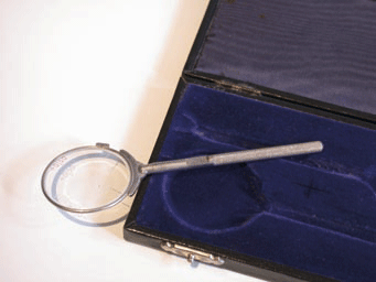 |
| The familiar cross cylinder |
When working with infinity charts you need no adjustment of the sphere for the testing distance. Direct 6m charts should, in theory, cause over-plussing of 0.167D, which is why pre-presbyopes like to be left on the green.
Presbyopes usually have smaller pupils, giving greater tolerance to blur, and appreciate the slight boost to their mid-distance vision that the extra quarter-dioptre gives.
Projection charts with a 3m distance will cause over-plussing of 0.33D, so it is customary to add -0.25DS to the distance portion of the subjective findings before prescribing. In the case of a presbyope, any extra minus on the distance prescription should be offset by adding +0.25DS to the reading addition.
|
This article has been adapted from Routine Eye Examination by Bill Harvey and Andy Franklin, part of the new Eye Essentials series. ORDER YOUR COPY |