
Huvitz: HOCT
The HOCT from Huvitz, distributed by Mainline in the UK, is said to feature high-speed and high-quality scanning (68,000 angiography A-scans per second) and 3D rendering of retina and macula thickness/separation. The device combines OCT, fundus camera, angiography (HOCT-1F/FA) and a personal computer. The web browser allows data to be analysed independently from the machine without the need to install specialist software. Detailed reports showing pathological structure and important data in an easy-to-read format can be viewed on screen or printed. The device is said to allow for quick capture of a wide area scan (12x9mm), covering both the macula and optic disc in one go to analyse thickness maps from retinal nerve fibre layer, ganglion cell layer and retinal pigment epithelium layers.
Optopol: Revo
UK distributor BIB describes the latest range of Optopol Revo OCTs as affordable, fast, compact and fully automated, making it a leading choice for primary care screening. BIB says the device produces the highest quality tomograms, which provides exacting and repeatable results for accurate progression analysis and monitoring over time. Referral data can be sent in the newstandardised digital imaging and communications in medicine format.
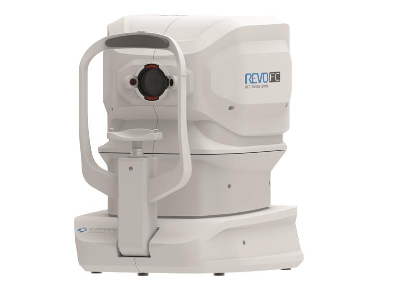
Optopol is said to boast the fastest OCT available at 130,000 A-scans and has introduced more features for improved functionality. These include an improved 12.3 megapixel colour camera for true colour fundus imaging, an artificial intelligence denoise algorithm for improved tomogram visualisation, a wider full scan range of 16mm (12x12), and full visualisation of the anterior chamber and lens (3-16mm) with no adaptor required. Additional features include biometry providing axial length for myopia management, topography of front and back surface, angiography, real-time eye tracking, and structure and function analysis incorporating visual field results for improved glaucoma monitoring and quantification. The supplier also notes that unlimited review licences are supplied free of charge.
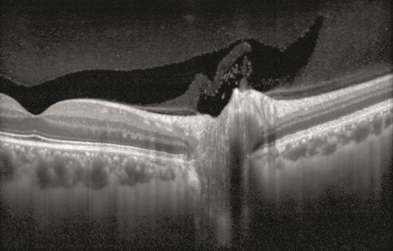
Topcon: DRI Triton OCT
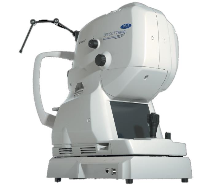
The Maestro2 provides colour fundus photography and automated non-invasive OCT angiography in one compact instrument. Topcon said with a single touch, the Maestro2 automatically performs alignment, focus, optimisation and capture and displays an instant report. Even less-experienced operators can capture a 12x9mm widefield OCT scan in seconds, encompassing the macula and optic disc. Analysis tools, including the ‘hood report’, support visual field decisions and in full in first entry then OCT-A, allowing for careful monitoring. This year, Topcon has also added PixelSmart technology to its Triton Swept Source OCT, which ‘reduces speckle noise and improves contrast, further enhancing 3D scan quality’. The DRI Triton combines swept-source OCT technology with multimodal fundus imaging. It has a scanning speed of 100,000 A-scans per second, producing clear B-scans, while an invisible 1,050 wavelength visualises the deepest layers of the eye in a single image.
Grafton: Mocean 3000 SLO OCT
According to distributor Grafton, the Mocean 3000 SLO OCT uses a 36,000 A-scans per second scanning speed to enable high-resolution cross-sectional images with minimised speckle noise for powerful diagnostics. The device simultaneously acquires OCT images and 47 degree fundus images using SLO technology, providing a real-time overview of the retina that allows easy localisation of lesion areas before acquisition. The Mocean 3000 features three-in-one scan capability, comprising macula, disc and cornea, while using eye tracking for increased scan repeatability. The high-speed OCT and SLO camera features are said to enable detailed >12mm scan width and the intuitive software is built to provide specifically what is needed for referrals and treatment decision-making. The device provides non-invasive, high-speed scanning and multiple patient contact adjustments for maximum patient comfort.
Optovue: iFusion80
Optovue’s iFusion80 combines the iVue80 SD-OCT system with the high-resolution iCam12 12 megapixel fundus camera. The system now offers 80,000 A-scans per second – three times faster than the original model – offering improved efficiency and enhanced image quality. Increased scan patterns and a higher resolution offer clinicians increased confidence in the diagnosis of pathology. The device also allows an exclusive iWellness scan, which checks for both retinal disease and glaucoma, offering the patient further peace of mind. The system boasts a comprehensive range of anterior scans, including epithelial thickness mapping for the measurement of dry eye and vault mapping for those practices offering scleral lens fitting. The cost-effective modular system allows the user to purchase the iVue80 and the iCam12 individually, with the option to upgrade at any time, benefiting practices with smaller budgets.
Heidelberg Engineering: Spectralis
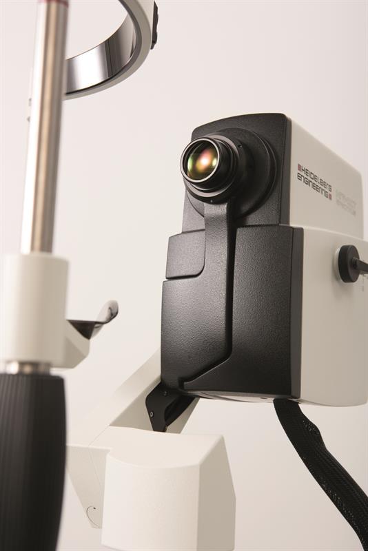
The Spectralis from Heidelberg is a multimodality imaging platform with an expandable design that lets it grow as new technology emerges. This is said to make it a safer investment that will future-proof your practice for years to come.
Heidelberg says the Spectralis employs special technologies in order to achieve consistent, high-quality results that provide the optometrist with the information they need to manage their patients with confidence. Next generation fundus imaging with confocal scanning laser ophthalmoscopy makes high-resolution imaging possible, even through cataracts and undilated pupils. This is combined with patented TruTrack Active Eye Tracking, which actively tracks the eye in real-time throughout image capture to mitigate the effects of eye motion and blinks. TruTrack makes precise, automated follow-up OCT scanning possible with measurement reproducibility to within one micron.
Spectralis provides the optometrist with the tools they need to improve patient care by enabling them to detect small changes in the retina, accurately monitor these changes over time, and refer patients for treatment at the earliest opportunity.
Optos: Monaco
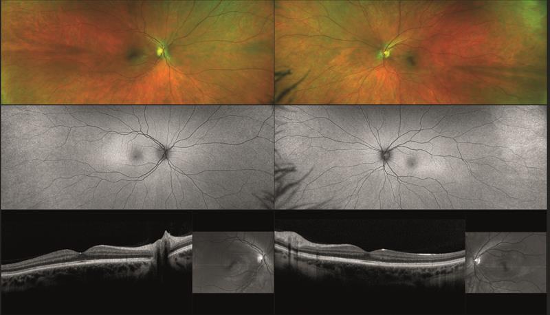
A pioneer in ultra-widefield retinal imaging, Optos is said to enable eye care professionals to discover, diagnose, document and treat ocular pathology. Monaco, an ultra-widefield retinal imaging device with integrated OCT, produces a 200 degree single-capture optomap image in less than half a second and also provides cross-sectional 40 degree OCT views of retinal structures. According to Optos, the Monaco device can capture a six-image, multimodal overview of both eyes in as little as 90 seconds. Visualising multiple image modalities at the same time enables the practitioner to detect pathology in the various views. This quick overview is also said to help improve clinic flow as the practitioner can see everything at once.
Nidek: Retina Scan Duo 2
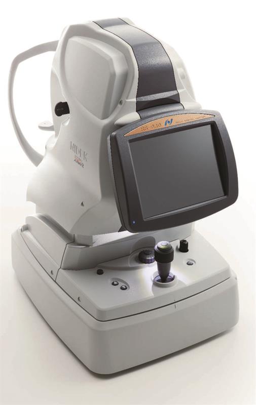
The updated Nidek Retina Scan Duo 2 (RSD 2) combined high-resolution OCT and colour fundus camera (12 megapixel) benefits from high-resolution 6x6mm macula and disc OCT-A with vessel density and perfusion density displays. Available from December, it features a retina map scan, incorporating macula and disc scans and colour fundus in one rapidly captured scan with an intuitive clinical data display. An automatic B-scan reduces noise to maximise scan resolution. The one-touch fully automated device provides exports for Opera and the Cues platform, sending OCT scans to the NHS referral service. An anterior module is included, while Navis 1.11 software provides a simple follow-up with progression analysis and trend prediction for advanced diagnostic understanding. The RSD 2 produces 70,000 A-scans per second and offers nine retinal scan patterns and four anterior scan patterns.