OPTICIAN looks at a recent addition to the simple yet effective card based methods of vision screening
All eye care practitioners and increasing numbers of patients are familiar with the Amsler chart for screening and monitoring of central vision changes related to macular disease. Its very simplicity of use and interpretation has led to its widespread popularity, but some concerns have been raised regarding its sensitivity.
The conventional black and white Amsler test grid fails to detect 87 per cent of early and small size (less than six degrees) macular field defects and 45 per cent of all macular field defects. Only one out of every three cases with wet macular degeneration and choroidal neovascularisation may be detected by the conventional grid. As the new treatment options become available for the treatment of choroidal neovascularisation, the earliest possible detection of such lesions is now even more important than before for timely delivery of vision-saving care. The red-on-black version of the Amsler grid is too difficult to be seen (very low contrast) by many patients and creates an unacceptably high false alarm rate.1,2
US optometrist Dr Erik Mutlukan has developed three further cards, the Ixmus Color Visual Field Test Cards, which exploit contrast and colour to enhance their sensitivity, compared with traditional Amsler cards, when screening central vision. Each is designed to be viewed monocularly at 30cm in good light and with fixation maintained upon a central target on the cards.
Card1
Card 1
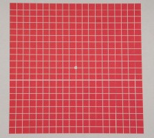
Card 1 is similar to a reverse version of the colour Amsler in that it has red squares on a white background, each square subtending about 1 degree of arc when viewed at 30cm (assessing therefore 10 degrees either side of fixation in total). With adequate fixation, it should be sensitive enough to detect central vision changes, particularly those related to toxic effects or early optic nerve defects. The designer recommends this card is used to retest any questionable results arising from cards 2 and 3.
Card 2
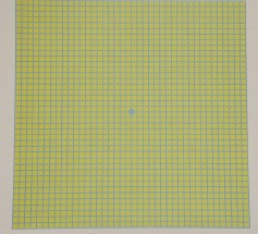
Card 2
Unlike the high contrast of card 1, this low-contrast card has a blue line grid over a yellow background with each square subtending half a degree at the retina. This much-lower contrast target should offer much greater sensitivity when screening for early toxic or other maculopathy.
Interestingly, the manufacturers state that it is useful for screening for senile lens sclerosis, though it is questionable this would offer anything more useful than direct viewing of the lens or contrast screening.
Card 3
Card 3
This is another low-contrast chart (Figure 3) with a mauve grid over a red background, leaving checks of 1 degree subtense for a 30cm viewing distance. It is surrounded by eight large red circles, two on each side. The grid provides a reasonably sensitive chart for early glaucomatous scotoma, while the larger red dots around the outside detect any gross neurological defect. It is claimed that the red-dots perimetry card is 90 per cent sensitive3 in detecting optic nerve and neuro-ophthalmic field defects and that is further enhanced by the addition of the grid centrally.
It certainly cannot be disputed that the test takes a far lot less time to administer than a gross perimetry with a bead on a stick.
Though simplistic in design, the grids seem readily accepted by patients and should prove useful where Amsler readings are inconclusive or inadequate.
References
1 Validity and interpretation of Amsler grid reports. Arch Ophthalmol, 1993; 111: 776-780.
2 The Amsler Chart is of doubtful value in retinal screening for early laser therapy of subretinal membranes. The West London Survey. Eye, 2004; 18, 503-508.
3 Red square test for visual field screening. A sensitive and simple bedside test. Acta Ophthalmologica (Copenhagen), 1994 Dec;72 (6):683-7.
For more information see www.ixm.us