Optopol: Revo OCT
Optopol’s new ‘Structure and Function’ analysis software combines the results of its Revo OCT (Structure) with the results of its PTS 920 Visual Field Analyser (Function) for the improved diagnosis and monitoring of major eye conditions, particularly chronic open angle glaucoma (COAG). The Revo NX 130, launched this year, delivers an impressive 130,000 A scans per second – ‘the fastest on the market’, according to Optopol. As with all Revo OCTs, including the Revo 60, Revo 80 and Revo FC (with colour fundus imaging), the Revo NX 130 provides posterior and anterior scans and optional biometry, angio and topography software. Fully automated or manual modes are available while a voice guides the patient through the scan process. The devices can be operated remotely from another consulting room or even off-site, enabling professionals to ensure social distancing and shield vulnerable patients. Downloadable marketing materials include patient leaflets, adverts, posters, press releases and phone scripts.
Price: £35,995
01438 740823
Heidelberg Engineering: Spectralis OCT
The Spectralis OCT features ‘TruTrack Active Eye Tracking’ patented technology, using a second laser beam to track the eye during scanning and avoid motion artifact. TruTrack averages up to 100 OCT B-scans, and produces images of such high resolution that clinicians can segment each layer of the retina. These images, along with thickness maps, are clearly presented in the software for analysis. The confocal optics and invisible infrared light source are comfortable for patients and pupil dilation is not needed. Auto rescan technology automatically repositions the OCT scan in the same anatomic location, detecting changes as small as one micron so that clinicians can confidently monitor disease progression over time. More than ‘200 NHS hospitals use Spectralis equipment’, enabling professionals who also use the devices to easily share results and build relationships with ophthalmologists. Regularly updated hardware and software most recently includes a reference database for retinal, RNFL, GCL and IPL thickness and the ‘Hood Glaucoma Report’.
Price: £39,995
01442 502330
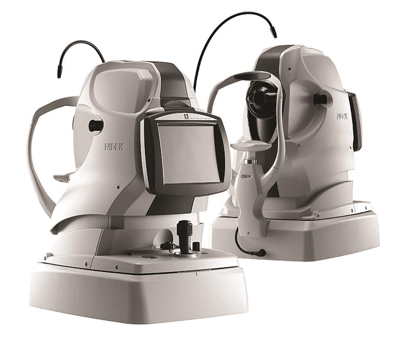
Nidek: Retina Scan Duo OCT
The compact, touch-screen Retina Scan Duo OCT from Nidek combines high resolution OCT and a full colour 12 megapixel fundus camera with auto fluorescence (FAF) capability.
With fully automated auto alignment, tracking, focus and capture for both OCT, fundus photography and FAF, the one-touch operation system can be easily delegated to other staff members, allowing more time to analyse and discuss results with patients.
The device provides exports for Opera and the Covid-19 Urgent Eyecare Services platform, sending OCT scans to the NHS referral service. An anterior module is included as standard and it provides a simple follow up with progression analysis and trend prediction for advanced diagnostic understanding.
The Retina Scan Duo OCT produces 53,000 regular scans, 26,500 fine scans and 13,250 ultra fine scans per second with a resolution of four microns. It offers eight retinal scan patterns and four anterior scan patterns, including pachymetry and anterior angle assessment. The company offers customers training, education and marketing support.
Price on enquiry
0808 123 2020
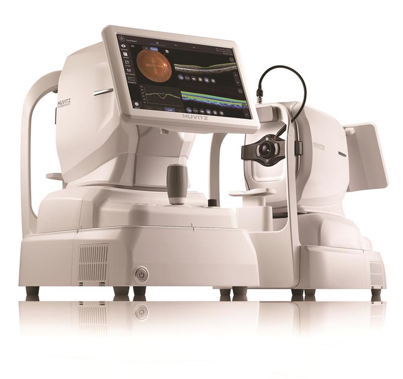
Huvitz: HOCT
The Huvitz HOCT is a fully integrated OCT, fundus camera (HOCT-1F and 1FA models) and PC all-in-one device, offering high speed and high quality scanning plus OCT angiography in the HOCT-1FA model. The device has a 12mm by 9mm scanning area and produces a 12 megapixel colour fundus image, enabling professionals to quickly capture and view the retina in a single scan. The all-in-one design, has an optional add-on anterior module and features an intuitive software interface, auto-tracking to keep the device in alignment and an ‘auto shoot’ function. The anterior segment module enables clinicians to measure and analyse corneal thickness, anterior chamber angles and 3D image. A corneal thickness map can identify corneal irregularity and thinnest point. Professionals can tailor reports to display a quick summary or more complex evaluation. Data can be viewed remotely and images shared with patients via a web browser system.
Price: from £342.60 per month
0121 458 6800
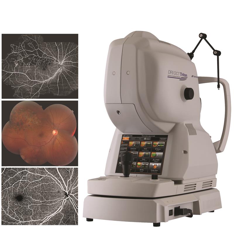
Topcon: DRI Triton OCT
Topcon’s DRI Triton OCT with combined anterior and posterior swept source OCT incorporates full colour high resolution fundus photography and FA and FAF imaging (Plus only). With a scanning speed of 100,000 A-scans per second, the Triton brings more scans for a single B-scan image and provides improved quality and efficiency. The 12mm B-scan covers both the macular area and the optic disc. A high penetration optimised long wavelength scanning light (1,050nm) visualises deep layers of the eye, such as choroid and sclera, in a single scan. The invisible wavelength prevents patients from being distracted, leading to fewer movement artifacts and increased repeatability. SmartTrack technology also follows the fixation movement, detects blinking and compensates for patient involuntary eye movements by delivering a follow-up scan in precisely the same anatomical location. The device’s combination scan is convenient and time-efficient for practitioners and patients, offering macular and RFNL plus macular and disc analysis in one overview.
Price on enquiry
01635 551120
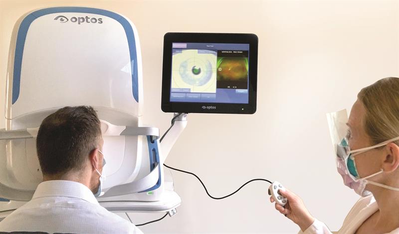
Optos: Monaco and Silverstone
Optos, known for its 200 degree single capture ultra-widefield optomap image, has introduced Monaco and Silverstone retinal imagine systems with integrated OCT.
These widefield two-in-one desktop retinal imaging systems, produce an optomap image in less than half a second as well as cross-sectional OCT views of retinal structures. Monaco provides cross-sectional 40 degree OCT views of retinal structures, and enables a rapid multi-modality capture, featuring colour, autofluorescence, and OCT scans, for both eyes, in two minutes. Silverstone, meanwhile, combines optomap imaging with integrated, image-guided, swept source OCT. The Silverstone produces a single capture optomap image with guided OCT allowing advanced OCT imaging anywhere across the retina, from posterior pole to far periphery. All Optos images are quickly acquired from a safe distance via a hand controller or tablet and images can also be reviewed remotely, ensuring patient safety and social distancing.
Price on enquiry
01383 843300
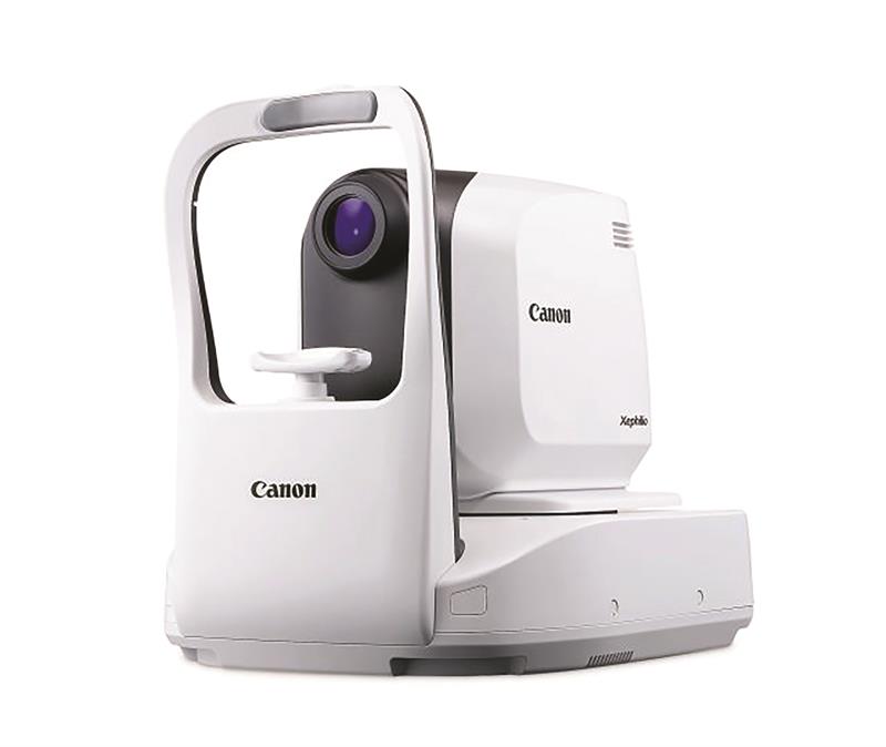
Canon: Xephilio OCT-A1
In three clicks of a mouse, the Canon Xephilio OCT-A1 can carry out patient scans a few metres away, from another room or even remotely, enabling practitioners to ensure safe social distancing. The Xephilio OCT-A1 system, comprising the OCT device, RX capture software, computer and LCD monitor, has a scanning speed of 70,000 scans per second with an optical depth resolution of three microns. Integrated scanning laser ophthalmic real-time retinal tracking technology automatically retains the scan position and protocol for each patient, eliminating the need for manual adjustment. The OCT averages up to 200 scans and offers specific scan modes for vitreous and choroid imaging with a scan width of up to 13mm. It can automatically detect and distinguish 10 layers of the retina and features 10 fixed, freely programmable exam pre-sets, comparing the results of five examinations arranged in a time sequence. Fundus images taken on an optional retinal camera can also be integrated into the OCT examination.
Price: £55,000
01494 775 811
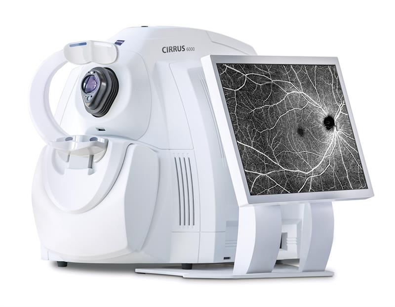
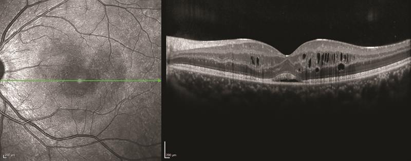
Zeiss: Cirrus 6000
Cirrus 6000 is the latest addition to Zeiss’ portfolio of OCT devices, offering increased efficiency and improved imaging detail. At 100,000 scans per second, the Zeiss Cirrus 6000 enables users to produce an image of a larger field of view of up to 12mm in a single scan. The device captures high-definition OCT and OCT angiography (OCTA) scans, revealing the finer microvascular details of the retina and enabling detailed insight into a patient’s condition.
The device also features a ‘Cirrus Wellness Report’ – designed to help educate patients. The simple and comprehensive report displays both macular and optic nerve head information compared to normative data on one convenient page. This allows clinicians to do a quick but thorough patient evaluation, ensuring optimal patient throughput and high practice efficiency. Other OCT devices in the range include the Cirrus HD-OCT 500, Cirrus HD-OCT 5000, Cirrus photo 600 and Cirrus photo 800.
Price on enquiry
01223 401500