Features

A case of dry eye neurosensory pain

Clinical Practice
Optometrist Dr Keyur Patel presents another challenging case from his practice, this time concerning a patient with a long history of ocular surface disease

Initial presentation

A 30-year-old male presented for a scleral contact lens fitting as part of the management of his long standing ‘dry eye’ problems. Scleral contact lenses had been suggested as a potential management option at his last eye examination by a colleague at another practice.

At present his only management was the use of Systane Gel Drops as needed, which in practice might be as frequent as every 10 minutes.

Key points from his history included the following;

  • At the age of 14, he had suddenly become very sensitive to light and was ‘squinting’ all the time. In his own words, the eyes looked ‘glazed and eyelids felt heavy. There was lots of sleep on my eye lashes. This affected my school life, with accusations of drug abuse from everyone, even teachers.’
  • He visited the doctors at this time (could not recall what was said), but bought some eye drops which did not help. After that he just ‘accepted this was how his eyes were’ and did nothing for four years.
  • At about 18 years old, he was referred to Milton Keynes University Hospital (by his GP) where he was diagnosed with blepharitis and seen for ‘a couple of visits.’ He was asked to undertake lid hygiene and warm compress procedure but found that this made his symptoms worse. He stopped after a few attempts.
  • Meibomian gland expression was tried, but ‘nothing came out.’
  • Punctal plugs were inserted at the hospital which helped to a degree but made the eyes very watery so were subsequently removed.
  • Having ‘tried pretty much every drop on the market, I came across Systane gel drops which is the only drop that provides relief. They have turned my life around and given me my independence back.’ He reported complete reliance on Systane and needs to instil every 30 minutes throughout the day –sometimes longer and sometimes more frequently.
  • In 2012, due to constant redness, went to the USA to have I - Bright treatment (see discussion later).
  • At this presentation, the patient stated ‘the eyes don't hurt - they are just dry and irritated.’

Clinical assessment

Initial clinical findings are summarised in table 1.

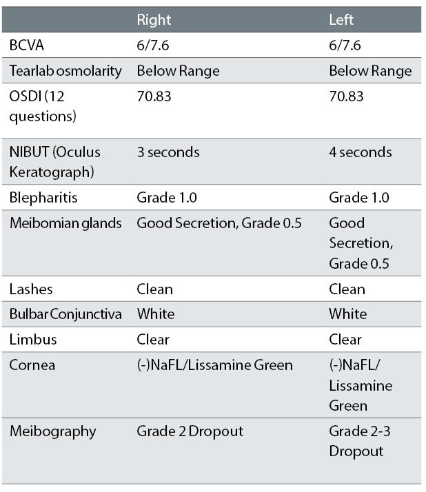
Table 1: Clinical findings at first presentation to author

Figures 1 and 2 show the lids after treatment with Blephex, an in-office procedure that uses a hand-held device (figure 3) that spins a medical grade micro-sponge along the edge of the eyelids and lashes, removing scurf and debris and exfoliating the eyelids.17

Figure 2: Left lids after Blephex treatment

Figure 3: The Blephex lid treatment in operation

The bulbar conjunctiva (figures 4 and 5) and ocular surface viewed with blue light and fluorescein (figures 6 and 7) were unremarkable.

Figure 4: Right bulbar conjunctiva

Figure 5: Left bulbar conjunctiva

Corneoscleral topography was undertaken using the Eaglet Eye ESP, capable of mapping an area covering the entire cornea and extending onto the sclera (figures 8 and 9). Meibography images taken with the Topcon Triton using the anterior segment lens are shown in figures 10 and 11.

Figure 6: Right NaFl appearance

Management

The patient was initially treated with Blephex in practice to clean the lids, followed by an Ocusoft Swabstix (single use swabs for the lid containing 50% tea tree oil, 40% sea buckthorn oil and 10% caprylic acid).16

Figure 7: Left NaFl appearance

Based on the appearance of the eye at presentation, and the patient’s refraction (he had low grade myopia with minimal astigmatism), the use of a daily disposable contact lens (Dailies Total 1, Alcon) was considered as an alternative option to the more complicated scleral lens fitting.

Figure 8: Eaglet Eye corneoscleral topography map for the right eye

Dailies Total 1 (DT1) contact lenses were inserted into both eyes. Initially the patient reported no difference in sensation, but when the lenses were replaced with trial scleral lenses (Zen Lens, Bausch and Lomb), he reported an immediate reduction in comfort and increased sensation. The initial Zen lens was selected using the fitting guide based on the HVID. He definitively preferred the comfort of the DT1.

Figure 9: Eaglet Eye corneoscleral topography map for the left eye

Plan

The patient was carefully tutored about the importance and maintenance of lid hygiene, including the regular use of warm compresses as well as improved hydration and tailored nutrition. He was carefully instructed in the application and removal of contact lenses and issued full instructions (both as a hard copy and in electronic form via Captiv8 Share) along with a trial supply of the lenses. He was also issued with a preservative-free topical lubricant to be used in conjunction with the contact lenses to be used as required.

Figure 10: Right eye meibography images (Topcon Triton)

A follow-up appointment was set for seven to 10 days later.

Follow up

The patient attended after one week and reported that his eyes felt much better with the contact lenses. He was still using the Systane Gel tears (as the preservative-free topical lubricant was not as effective), but he was ‘not as reliant' on this as previously, typically using the drops every hour. The contact lenses had been worn for a maximum of 15 hours, and at presentation for six hours wear. He was happy with the vision, the lenses fitted well, and there was no disturbance of the cornea or anterior surface.

Figure 11: Left eye meibography images (Topcon Triton)

We had a discussion about the use of preserved artificial tears and contact lenses (and potential risks), and it was recommended to try Systane Ultra (preservative-free) drops as alternative. He was advised to continue with the lid hygiene and warm compresses and reminded of the importance of maintaining these treatments.

Discussion

‘Dry eye is a multifactorial disease of the ocular surface characterized by a loss of homeostasis of the tear film, and accompanied by ocular symptoms, in which tear film instability and hyperosmolarity, ocular surface inflammation and damage, and neuro-sensory abnormalities play etiologocal roles’ as defined by the TFOS DEW II Definition and Classification Report.¹

A diagnosis of ‘dry eye disease’ (DED) is given to someone who has a positive symptomology questionnaire score (DEQ 5 or OSDI), and at least one of the following;

  • Non-invasive breakup time of less than 10 seconds
  • Osmolority of >308 mOsm/L (or an asymmetry of >8mOsm/L)
  • Ocular surface staining (>5 corneal spots or > 9 conjunctival spots or lid margin staining ≥ 2mm length and 25% width) 1

The DED can then be classified as either aqueous deficient dry eye, mucous deficient dry eye or mixed dry eye.

The clinical decision-making algorithm1 categorises patients as

  • Signs without symptoms
  • Neurotrophic conditions
  • Other ocular surface disease (differential diagnosis)
  • Symptoms without signs: pre-clinical state
  • Neuropathic pain (non-OSD) – this needs to be referred for pain management.

The aim of management is to restore homeostasis of the tear film.

Restoration of tear film homeostasis is a multimodal ongoing form of management with treatment starting with patient education regarding DED,2,7,9 then moving forward with the following range of options;

  • Modifying environment
  • Discussing nutrition
  • Managing the lid margins and MGD (Demodex treatment if necessary)
  • Artificial tear supplement
  • Punctal occlusion
  • Moisture glasses
  • Topical prescribed medications
  • Oral prescribed medications
  • Therapeutic contact lenses
  • Oral secretagogues
  • Autologous/allogenic serum eye drops
  • Amniotic membrane
  • Surgical intervention (such as tarsorrhaphy)

The symptoms in this case were severe (based on OSDI), but this does not correlate with presenting signs, which suggests a neurosensory element to the DED. The first line of treatment was to treat the blepharitis (Blephex), followed with a Demodex swab and an explanation of the importance of maintaining good lid hygiene.

The cornea is the most innervated tissue in the body, 7,9 with approximately 7000 nerve terminals per square millimetre 7,9 which is 300 to 600 times greater concentration than in the skin, and 20 to 40 times that of dental pulp.9

The cornea and anterior bulbar conjunctiva are supplied by the nasociliary nerve, a branch if the ophthalmic division of the trigeminal nerve. 3,5,6,7,9 The supratrochlear, supraorbital, infratrochlear and lacrimal branches of the ophthalmic nerve (as well as the infraorbital nerve of the maxillary nerve) supply the innervation to the remaining bulbar conjunctiva, the entire palpebral conjunctiva, and the skin around the eyelid margin. The cell bodies are in the ophthalmic and maxillary region of the trigeminal ganglion.3

The corneal nerves enter the corneal epithelium and Bowman's layer radially (at about 1mm from the corneoscleral limbus) and lose myelination at the site of entry.3,7,9 Some nerves then terminate as free nerve endings, others from anatomical association with stromal keratocytes, while many nerves penetrate Bowman's layer to rest under the basal epithelial layer to form a sub-basal nerve plexus. The nerve plexus then gives rise to superficial intraepithelial terminals which end as bulbous free nerve endings that contain nociceptors.7

Pain disorders can be divided into nociceptor and neuropathic diseases.5 Nociceptor pain is usually due to tissue damage or inflammation and usually transient, whereas neuropathic pain is defined as ‘pain arising as a direct consequence of a lesion or disease affecting the somatosensory system.’ 3,4,5,6

Corneal nociceptors can be divided into polymodal, cold thermo and mechanical receptors and the differences are summarised in table 2.

Table 2: Characteristics of corneal nociceptors

Corneal receptors and function

Conjunctival nerves are similar to those of the cornea,3 but less sensitive. The eyelid margin nerves have a larger proportion of nerve endings which are designed to detect very low level mechanical stimuli.3 It has been shown that sensory nerves are structurally and functionally altered in dry eye disease.³ The density and location of corneal sensory nerves makes them particularly susceptible to adverse environmental conditions, trauma and disease.

Tear deficiency can leave the cornea exposed to environmental factors and increased evaporation can cause increased osmolarity. A wide variety of factors can affect corneal sensory nerves, but it still not known which single or combination of factors, or duration of challenge is needed to cause sensitisation.3

These factors cause local inflammation, which can lead to long and short term genetic and molecular changes that modify the electrophysiological characteristics of the peripheral terminals.3

Inflammatory environments can lead to peripheral sensitisation. Peripheral sensitisation can cause ectopic firing of nerves, the spontaneous generation of action potentials, aberrantly enhanced conduction of signals, increased excitatory neurotransmitter release, nerve sprouting, and the re-wiring and conversion of non-nociceptive sensory afferent nerves into fibres exhibiting a nociceptive phenotype.6,7 The patient can be sensitive to stimuli that would not normally cause pain, such as photo allodynia, and may develop a higher pain response.6,7, 8

Peripheral sensitisation can be reversed by treating the causes of inflammation and hyperosmolarity.5 If, however, ocular surface damage and inflammation continue, changes can occur within the central nervous system.5 When changes occurs at the level of the central nervous system, the level of pain does not correspond to initiating stimulus or mirror the signs.5,6,8

Neuropathic pain tends to be chronic and can be associated with any part of the body,4,5,6,10 but it is not caused by the pathophysiology of dry eye disease.3 Neurosensory dysfunction is recognised in dry eye disease and may account for the often-poor association between signs and symptoms.³ The descriptive terms ‘burning’, ‘shooting}, ‘pins and needles’ and ‘itching’ appear to be specific to neuropathic pain.6,7

Dry eye symptoms are often described by patients with chronic pain conditions,6 as well as those diagnosed with depression and anxiety. Patients with neurosensory changes benefit from treatment of the ocular surface disease with protective and anti-inflammatory agents, and by treating their central ocular sensory dysfunction with anti-neuropathy drugs.4,5 The mechanism of dry eye symptoms associated with comorbidities of depression and anxiety are not yet fully understood.5

I – Bright surgery is similar to conjunctivoplasty, promising ‘whiter and brighter eyes’. It is a modified form of conjunctivoplasty and is often used for treating pinguecula, pterygia, conjunctivochalasis and other disorders of the ocular surface. The procedure involves local conjunctival resection of those areas that have excessive or dilated vessels, hyperplastic growths (such as pinguecula or pterygium) or dense melanosis.12

The treatment goal for the neurosensory element of dry is to reduce the signalling from the corneal nerves. This is achieved by the following; 6,7

  • Lubrication – diluting inflammatory markers, decrease osmolarity
  • Punctal plugs – which increase the tear reservoir
  • Anti-inflammatories – there is a link between inflammation and pain amplification by nerve sensitisation
  • Autologous serums – trophic factors within the serum promote health and regeneration of the corneal stroma and sensory nerves
  • Anti-convulsant therapy
  • Anti-depressants
  • Analgesics
  • Soft contact lenses – these may shield sensitised corneal receptors from environmental stimuli
  • Acupuncture – stimulates endogenous opioid mechanisms
  • Vigorous exercise – increases brain neurotrophic factors and thereby contributes to neuroplastcity and neuro-restoration
  • Anaesthetic testing – differentiates peripheral and central sensitisation.7

To reduce the level of inflammation along the lid margins and on the ocular surface in this case, first and foremost it was important to manage the blepharitis.

Demodex folliculorum is a microscopic mite that can only survive on the skin of people. Most people have D. folliculorum on their skin. Usually, the mites do not cause any harm. The entire life cycle of D. folliculorum takes 14 to 16 days Adult mites mate at the top of the hair follicle, near the skin surface. Eggs are deposited in the sebaceous gland inside the hair follicle. It takes seven days for the larva to develop into a mature adult, via two intervening nymph stages.

The adult lives for four to six days.19 Tea tree oil (TTO) is a natural product distilled from the leaf of Melaleuca alternifolia. Gao et al.12 showed its effectiveness in killing Demodex in vitro and in vivo and introduced a TTO lid scrubbing treatment which has proved to be very effective in decreasing Demodex on eyelashes. To maintain lid margin cleanliness, our patient was advised to maintain lid hygiene at home, twice a day with Ocusoft Oust Foam (which also contains tea tree oil).

Warm compresses were also recommended to reduce viscosity of the meibum allowing easier expression. Hyperkeratization (due to hormonal disturbances, toxic effects of medications or chemicals, breakdown of meibomian lipids or external factors),³³ will cause blockage of the ducts. This in turn is believed to cause increased pressure within the glands leading to atrophy of the meibum secreting acini and subsequently the whole gland (as imaged by meibography).33

Historically, preserved drops have not been recommended with contact lenses, which could be related to preservative associated transient hyperfluorescence (PATH). In Contact Lens Complications, Nathan Efron describes PATH as being seen typically between 30 mins to four hours after soft contact lens insertion and resolving after 6-8 hours. It is asymptomatic, and appears to lack future complications.32 He also writes that a true toxic reaction is usually pathological, symptomatic and may take several days to resolve. There do not appear to be any studies with regards to preserved drops and daily disposable wear.

Systane Gel Drops contain polyethylene glycol (PEG) 400 (0.4%) and propylene glycol (0.3%), with the active agent hydroxypropyl guar, and is preserved with polyquaternium-1 (0.001). Systane Gel Drops is formulated with a pH of 7.0 and a relatively low viscosity; once exposed to the higher ocular pH, it forms of a gel that helps to adhere to the ocular surface.25 Systane Ultra has similar formulation but is available in preservative-free dosing.22

Studies with regard to preservatives and the ocular surface have shown that benzalkonium chloride is cytotoxic and dose dependent. The immunological responses are more frequently of the delayed type (type IV hypersensitivity) than of the immediate type (type I).

Polyquad is a preservative derived from benzalkonium chloride of the quaternary ammonium class. It is considered less toxic to the corneo-conjunctival surface. Due to its large molecular size, it is not absorbed into hydrogel lenses and toxic and allergic reactions are rare.23

Systane Gel and Ultra use polyquaternium ammonium chloride (0.001%) and this had no discernible effects on cytokinetic movement or on the mitotic activity of the epithelial cells.30

With this knowledge, and appropriate education and follow up, it was felt Systane Gel could be used ‘off label’ with the daily disposable contact lens. We recommended that he try a unit dose variant, to try and eliminate as many potential inflammatory triggers as possible, whilst still keeping him comfortable.

The idea of a contact lens in the management of dry eye is to protect the corneal surface (and the corneal nerves),and help with dehydration. The contact lens of choice should have full coverage, medium to low water content (with minimal on-eye dehydration) and be frequently replaced.32

DT1 (Alcon) is made of delefilcon A. It has 'water gradient' design, with a core water content of 33% increasing to more than 80% at the surface. The high edge water content making it 'wettable and lubricious' with a Dk of 140.22,31

In practice, we have had an excellent response to this contact lens, especially with people would were reporting dryness symptoms. Our personal experience is consistent with that by Marx et al.24 in a small study of first time contact lens wearers.

Maisa et al.25 reported that DT1 lenses showed the same lubricity after 16 hours of wear as an unworn lens. It is known that the comfort of a lens has been shown to be linked to its coefficient of friction.21 Varikooty et al, 28 in a study of 100 patients, found that non-invasive tear breakup time was longest and wettability was greater with DT1.

Conclusion

An understanding of the neurosensory component of dry eye, along with its short and long-term consequences, is very useful when educating patients about the importance of compliance, and in aiding the practitioner when deciding how to treat.

The modern optometrist already has the skill set and most of the tools needed to diagnose and manage the majority of dry eye disease cases without the need for further referral.

As we learn more about dry eye disease, and the 'market' for it increases, many manufacturers are providing learning opportunities to increase knowledge at minimal if not zero cost to the practitioners. There are multiple articles, forums and discussion sites for practitioners to share and learn.

If you wish to run speciality clinics there are many additional tools that are available, and sometimes it is even possible to use the tools already in practice ‘off label’.

There are lots of products available by multiple manufacturers. Unfortunately, there is no ‘gold standard’ recommendation on any single course of action. To complicate matters, a product that works for one patient may not work for their twin. From our experience, Total Dailies 1 has had very positive response from the majority of our patients.

With so many permutations and variables, it can be hard for a practitioner let alone the patient, and there is much to gained from keeping treatment and management as simple as possible.

At this stage, with the patient presented here, the use of readily available, ‘off the shelf’ products has had a life changing effect.

Dr Keyur Patel is a therapeutic optometrist working in Northampton.

The author has no financial interests in Alcon, but would like to thank Jonathon Bench, Amrik Matharoo and Priya Chahal for their help with research.

References

1 Craig JP, Nichols KK, Akpek EK, et al. TFOS DEWS II Definition and Classification Report. The Ocular Surface 2017;15:276–83.

2 Jones L, Downie L, Korb D, et al. TFOS DEWS II management and therapy report. The Ocular Surface Published Online First: 1 July 2017. doi:10.1016/j.jtos.2017.05.00.

3 Belmonte C, Nichols JJ, Cox SM, et al. TFOS DEWS II pain and sensation report. Ocul Surf 2017;15:404–37.

4 Mass MBA MD, CM, FRCSC, FARVO, Perry Rosenthal, MD, CM, and James McLaughlin, PhD, Andover. Neuropathic Pain: The Artifice of Dry Eye. https://www.reviewofophthalmology.com/article/neuropathic-pain-the-artifice-of-dry-eye.

5 Galor A, Levitt RC, Felix ER, et al. Neuropathic ocular pain: an important yet underevaluated feature of dry eye. Eye 2015;29:301–12.

6 Galor A, Moein H-R, Lee C, et al. Neuropathic pain and dry eye. The Ocular Surface 2018;16:31–44.

7 Goyal S, Hamrah P. Understanding Neuropathic Corneal Pain-Gaps and Current Therapeutic Approaches. Semin Ophthalmol 2016;31:59–70.

8 Jacobs DS. Diagnosis and Treatment of Ocular Pain: the Ophthalmologist’s Perspective. Current Ophthalmology Reports 2017;5:271–5.

9 Ocular Neuropathic Pain — EyeWiki. http://eyewiki.aao.org/Ocular_Neuropathic_Pain

10 Galor A, Zlotcavitch L, Walter SD, et al. Dry eye symptom severity and persistence are associated with symptoms of neuropathic pain. British Journal of Ophthalmology 2015;99:665–8.

11 Satitpitakul V, Kheirkhah A, Crnej A, et al. Determinants of Ocular Pain Severity in Patients With Dry Eye Disease. American Journal of Ophthalmology 2017;179:198–204.

12 I-BRITE Conjunctivoplasty for Improved Confidence — Boxer Wachler Vision Institute. http://www.boxerwachler.com/i-brite-eye-whitening.

13 O.D AGK OD,and Joseph W Sowka. Eye Whitening 90210. https://www.reviewofoptometry.com/article/eye-whitening-90210-27071

14 Benelli U. Systane lubricant eye drops in the management of ocular dryness. Clin Ophthalmol 2011;5:783–90.

15 InflammaDry | Quidel. https://www.quidel.com/immunoassays/inflammadry (accessed 25 Apr 2018).

16 OCuSOFT. OustTM Demodex SwabstixTM. http://www.ocusoft.com/oust-demodex-swabstix

17 BlephExTM: The First and Only Clinician Treatment for Blepharitis. http://www.blephexlids.co.uk/

18 Innovasium. TearLab. https://www.tearlab.com/

19 Demodex folliculorum. Wikipedia. 2018.https://en.wikipedia.org/w/index.php?title=Demodex_folliculorum&oldid =843456483

20 Gao Y-Y, Di Pascuale MA, Li W, et al. In vitro and in vivo killing of ocular Demodex by tea tree oil. Br J Ophthalmol 2005;89:1468–73.

21 Stewart — The friction coefficient of soft contact lens surf.pdf. http://openaccess.city.ac.uk/13791/1/Koller%2C%20%20A.W..pdf

22 The ACLM Contact Lens Year Book 2018. 2018th ed. The Association of Contact Lens Manufactures.

23 Coroi MC, Bungau S, Tit M. Preservativs from the eye drops and the ocular surface. Rom J Ophthalmol 2015;59:2–5.

24 Marx S, Lauenborg B, Kern JR. Performance evaluation of delefilcon a water gradient daily disposable contact lenses in first-time contact lens wearers. Cont Lens Anterior Eye Published Online First: 6 February 2018. doi:10.1016/j.clae.2017.12.019.

25 Maissa C, Nelson J, DeCenzo-Verbeten T, et al. Evaluation of the Lubricity of Delefilcon A Daily Disposable Contact Lenses (Dailies Total 1) After Wear.

26 Evaluation of the Lubricity of Dailies Total 1 Contact Lenses After Wear. https://www.aaopt.org/detail/knowledge-base-articl-after-wear.

27 Kern J, Rappon J, Bauman E, et al. Assessment of the relationship between contact lens coefficient of friction and subject lens comfort. Invest Ophthalmol Vis Sci 2013;54:494–494.

28 Varikooty J, Schulze MM, Dumbleton K, et al. Clinical Performance of Three Silicone Hydrogel Daily Disposable Lenses: Optometry and Vision Science 2015;92:301–11.

29 Cohen S, Martin A, Sall K. Evaluation of clinical outcomes in patients with dry eye disease using lubricant eye drops containing polyethylene glycol or carboxymethylcellulose. Clin Ophthalmol 2014;8:157–64.

30 Tripathi B, C Tripathi R, P Kolli S. Cytotoxicity of ophthalmic preservatives on human corneal epithelium. Lens and eye toxicity research 1992;9:361–75.

31 Contact Lens Spectrum ttps://www.clspectrum.com/content/alcon/10/cls_13_final_edit_pgs.pdf Efron N. Contact Lens Compications. 3rd ed. Elsevier.