![]()
![]()


Identify the changes within the MultiColor fundus image?
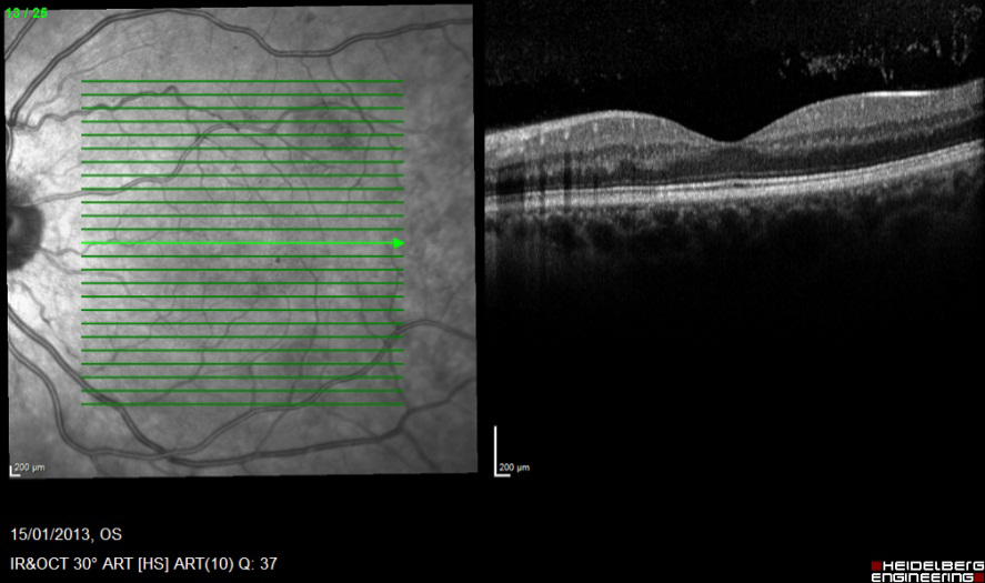
Multiple hard drusen (<63mm), multiple intermediate drusen (63-126 mm) pigment clumping and RPE atrophy. The changes are consistent with age related maculopathy, in which whitish-yellow drusen in the macular and hyper pigmentation and depigmentation of the RPE are seen. This is an early stage of macular degeneration AMD, which includes geographic atrophy or choroidal neovascularization in its presentation. Reticular drusen or reticular pseudodrusen have a poor prognosis for progression to AMD and important change to identify. These can be seen more clearly by using the blue/green enhanced display mode for MultiColor and fundus autofluorescence images (below).
Register now to continue reading
Thank you for visiting Optician Online. Register now to access up to 10 news and opinion articles a month.
Register
Already have an account? Sign in here