This is an uncommon, hereditary and usually bilateral macular dystrophy, in which yellow material (lipofuscin) resembling egg yolk (hence vitelliform) accumulates at the macula. 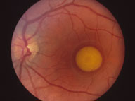
The inheritance pattern is autosomal dominant, with highly variable penetrance and expression. The genetic defect has been mapped to the VMD2/Bestrophin gene on chromosome 11.
The mechanism of accumulation of lipofuscin is controversial, and may relate to a defect in the photoreceptors or retinal pigment epithelium.
SYMPTOMS
Often asymptomatic. Patients may notice gradual reduction in visual acuity.
SIGNS
Signs are highly variable, even within affected families. The classical presentation occurs during the first or second decades, with the detection of a single, round lesion at the macula resembling an egg yolk.
Lesions are usually present in both eyes, located at the fovea, and measure approximately one to two disc diameters across.
Occasionally, unilateral, extrafoveal and multiple lesions occur. Despite the striking appearance of the lesions, visual acuity is initially normal or only slightly decreased in most cases.
The condition may be accompanied by micropsia. Central relative scotomas may be detected on careful perimetry.
Later in life, degeneration of the lesion often occurs, resulting in an irregular, 'scrambled egg' appearance. The yellow material within the lesion may then develop a fluid level ('pseudohypopyon'). These events are often accompanied by a substantial reduction in visual acuity and extension of scotomas.
Further degenerative change may result in scarring or macular atrophy, and occasionally in secondary choroidal neovascular membrane formation.
Register now to continue reading
Thank you for visiting Optician Online. Register now to access up to 10 news and opinion articles a month.
Register
Already have an account? Sign in here