
The following cases raise a number of points of interest:
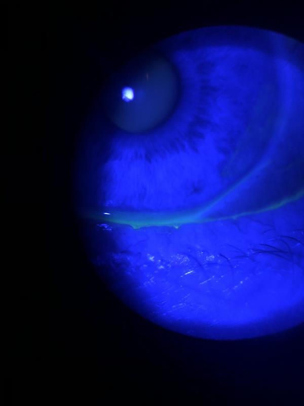 Figure 1a: Image of an eye under blue light after fluorescein instillation.
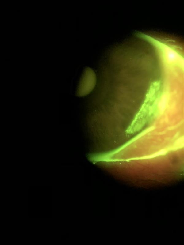 Figure 1b: Same eye with introduction of Wratten filter revealing peripheral corneal epithelial damage (Images: Beth Ralph)
Patient booked in to the dry eye clinic.
Presentation
Clinical findings
Figure 2: Video of fluorescein break-up time for left eye. At three seconds inferior desiccation visible, at five seconds break up spots visible
Figure 3: Video of fluorescein break-up time for right eye. Pre-blink appearance shows multiple sites of break-up, greater inferior desiccation than left and scattered desiccation stain over cornea. Moment of maximum blink showing clear lagophthalmos
Register now to continue reading
Thank you for visiting Optician Online. Register now to access up to 10 news and opinion articles a month.
Register
Already have an account? Sign in here