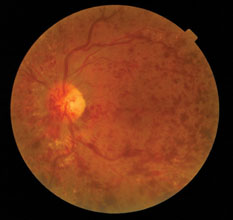
 Central retinal vein occlusion (CRVO) is a destructive condition with a dramatic ophthalmoscopic appearance: widespread haemorrhages in all quadrants of the retina. It is usually attributed to a thrombus in the central retinal vein, at the level of the lamina cribrosa within the optic disc. The vein at the arteriovenous crossing is also at risk of blockage, as a stiff-walled atheromatous artery may compress the lumen of the adjacent thin-walled vein, creating blood turbulence and thrombotic build-up.
CRVO is frequently associated with systemic diseases, particularly hypertension, arteriosclerosis, diabetes, and hypercoagulation or vasculitic disorders. CRVO also has an association with glaucoma, as raised intraocular pressure (IOP) may compress the central retinal vein.
There is a spectrum of disease in CRVO, in relation to the amount of venous obstruction, consequent stagnation of blood flow, retinal hypoxia and damage to the retinal capillary endothelial cells. The retinal capillary non-perfusion or closure (capillary ischaemia) is shown as hypofluorescence on fluorescein angiography. Damage to the capillary endothelial cells causes breaches in the blood–retina barrier, leading to the clinical signs of blood leakage from the vessels.
Register now to continue reading
Thank you for visiting Optician Online. Register now to access up to 10 news and opinion articles a month.
Register
Already have an account? Sign in here