 Choroidal haemangioma is a congenital, unilateral, benign tumour composed of endothelium-lined vascular channels. Two types exist:
Circumscribed: no association with systemic disease Diffuse: occurs in Sturge-Weber syndrome, characterised by facial port-wine stain (cutaneous haemangioma, or naevus flammeus), central nervous system haemangiomas and a high incidence of glaucoma (up to 70 per cent of cases).
Patients with choroidal haemangioma require regular review to enable timely detection and treatment of complications including cystic retinal degeneration and exudative retinal detachment.
Symptoms
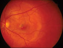
Circumscribed choroidal haemangiomas are usually diagnosed between the second to fourth decade when vision is affected by retinal detachment or cystic retinal degeneration. Symptoms of macular involvement include blurred central vision, micropsia and metamorphopsia. Diffuse choroidal haemangiomas are usually diagnosed in infancy during the assessment of Sturge-Weber syndrome.
Register now to continue reading
Thank you for visiting Optician Online. Register now to access up to 10 news and opinion articles a month.
Register
Already have an account? Sign in here