
Held in a conference centre just metres away from some of the Beach Boys’ favourite surfing haunts, the 2022 American Academy of Optometry (AAO), as always, offered a wide range of education and research from all areas of eye care practice. In this short feature, I will mention some of the key findings presented in the scientific programme, as selected by the Academy Board and their new president Dr Timothy McMahon (figure 1).
Glaucoma
Glaucoma remains one of the leading causes of irreversible blindness across the globe. Even with the advent of selective laser trabeculoplasty (SLT), at present, treatments are typically lifelong while decisions about treatments are based on the monitoring of disease progression. Because of the chronic nature of glaucoma, early and accurate detection and evaluation has major implications for a patient’s functional vision and quality of life.
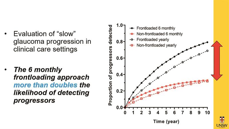
Dr Jack Pru and colleagues (University of New South Wales) described how current methods of functional assessment, such as threshold fields testing, often take many years before confirming disease progression. Modern threshold fields strategies, such as SITA Fast, have made it possible to undertake multiple visual fields tests at one single visit to a clinic with minimal cost. By adopting this ‘frontloading’ approach, with two fields assessments per visit, Chu has found disease progression is detected three to six visits sooner where disease progression is fast. For more typical, slowly progressing glaucoma, Chu has found that adopting a frontloading approach, with two fields tests every six months, nearly doubles the detection rate (figure 2). Indeed, the current ‘one fields test per visit’ approach misses around 70% of slowly-progressing glaucoma.
Figure 2: Two fields tests every six months doubles detection rates
Another way of predicting glaucoma progression has been looked at by Dr Michael Sullivan-Mee (New Mexico) and colleagues. Because of the variability in disease progression, being able to identify the more rapid progressors makes it easier to direct resources more appropriately. Recent studies have shown that any detected loss in the central 10 degrees of the 24-2 fields test is highly predictive of progression. However, there are only 12 test points in this area on the 24-2 test most of us use in everyday practice. The 10-2 test has 68 points within the central 10 degrees. Following on from two reliable 10-2 baseline tests, Mee and his team used 24-2 testing of 394 eyes every four to six months and monitored the rate of change over time (MD) and the likely progression (GPA analysis). What they found was that the most accurate predictor of glaucoma progression was the presence of defects found by the baseline 10-2 test. In fact, patients with 10-2 defects were found to progress at four times the rate. More impressive yet, the hazard analysis suggested that the presence of baseline 10-2 defects gave a 22 times greater risk of a further visual field loss.
Conclusions? We should consider more frequent fields testing and the use of the 10-2 strategy.
Contact Lenses
Paul Chamberlain (figure 3) is fast becoming a fixture at annual optometry conferences as his role heading up the ongoing MiSight 1 day clinical trial means he is able to report the latest results as they appear. At this year’s AAO, as the trial reaches its seventh year, the focus was on the response of fast myopia progressors to MiSight intervention. Despite great variability, the study has found that around 90% eyes showed significant slowing in eye growth (responders) and 10% little slowing (non-responders). Importantly, the fastest progressing eyes showed the most slow-down in progression with treatment, while the slower growing eyes stopped growing during treatment (figure 3).
Figure 3: Paul Chamberlain (inset) and results showing how the fastest growing eyes pre-treatment (red) showed the greatest response to MiSight treatment, the slowest (blue) the least
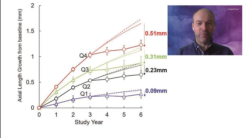
In other words, the eyes responded to treatment in proportion to their pre-treatment growth rate.
Anterior Eye
Another study from the University of New South Wales (UNSW) looked at the impact of probiotics and prebiotics on dry eye disease. There is increasing evidence that inflammatory processes throughout the body are influenced by the gut microbiome. So, might it be possible, by introducing oral probiotics and prebiotics, to alter the gut commensal bacteria, which modulate systemic inflammation and, therefore, minimise dry eye signs and symptoms? Using a double-masked, randomised controlled trial, 41 people with dye eye disease (DED) were assessed. After four months, the treatment group showed improvement with symptoms (p>0.001), not so the control group. Tear break-up time and meniscus height did not change for the treatment group but were reduced in the control. Early days yet, but this adds to the growing evidence for the usefulness of customised pre/probiotics in managing DED.
The use of artificial intelligence (AI) in the assessment of meibomian gland function is being studied at the University of California by Dr Meng Lin and colleagues. Meibography is familiar to most of us, but standardisation and quantification is proving difficult. AI, therefore, has been used to analyse image data to develop a prediction model of when problems are likely to occur. The model uses three inputs: the wide range of meibography phenotypes, the various morphology features of the glands and a wide range of clinical datasets. By so doing, the team have found that AI analysis of meibography images can predict expert clinician diagnoses for MGD (74% accuracy), blepharitis (73.7%) and aqueous deficiency (85.2% accuracy). Interestingly, the analysis can also predict age and ethnicity accurately. Conclusion? Meibography can be undertaken objectively and could soon be used as a biometric identifier, or ‘fingerprint’.
Dr Gauri Shrestha (again from UNSW) described his work looking at tear metabolomes (total number of metabolites) associated with culture-positive and negative microbial keratitis. Currently, only around 50% cultures from an active microbial keratitis are found positive through scrapes and these patients generally prove harder to treat than those with culture-negative keratitis. So, a biomarker that might help to diagnose culture-positive keratitis would be useful. It would appear that five tear metabolites differ in expression between culture-positive and negative keratitis and so might just be the biomarkers required.
Conclusion? A tear sampling test may replace a scrape and culture test for keratitis one day.
Systemic Health
Dr Lisa Zhu, from the Australian Centre for Eye Research, presented results from her study looking at the significance of retinal age as a biomarker for cardiovascular disease (CVD). CVD is a major killer and drain of health funding, even though many of the risk factors are modifiable (figure 4).
Figure 4: Many risk factors for CVD are modifiable
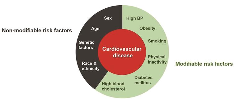
The highly vascularised nature of the retina makes it an ideal way to assess vascular health and, as Zhu’s team have confirmed, is an accurate way of assessing age. Therefore, by screening for signs of retinal ageing, it should be possible to predict those patients most likely to suffer from CVD and, therefore, most likely to benefit from interventions at an earlier stage.
Conclusion? A retinal photo may one day become as useful as other blood checks.
Visual Function
Vladimir Mamchik is a graduate optometry student already making waves in the world of colour vision. He has identified that current ways of helping those with colour deficiency, either by experimental viral vector genetic engineering or by the use of colour filters, are both limited in their scope. There is, therefore, a need for a safe, non-invasive correction method for the colour deficient. Mamchik has developed a system that, rather than changing the spectral composition of illuminating light, uses an active filter to control the wavelengths that stimulate the photoreceptors at high frequencies. This process has been found to introduce additional contrast between colours that previously could not be distinguished. The system has been shown to show significant improvements in colour discrimination in dichromats and monochromats. Expect to hear more about this when it moves beyond the laboratory.
Conclusion? There are some very smart young optometrists coming through the system.
