In the issue of September 19, Bill Harvey described the current grading nomenclature for diabetic
retinopathy and challenged readers to test their grading skills by classifying a series of images. Here
he suggests grades for each of the 12 presentations.
retinopathy and challenged readers to test their grading skills by classifying a series of images. Here
he suggests grades for each of the 12 presentations.
1 R1 M0. This retina shows a few scattered dot haemorrhages and possible microaneurysms, all well away from the macular area.
2
R1 M0. This retina shows some blot haemorrhages away from the macula. Note also the flame haemorrhage on the superior temporal branch vasculature, indicative of branch venous occlusion.
3
R1 M1. This retina shows more extensive non-proliferative changes with dot and blot haemorrhages spread throughout the posterior pole. There are also exudates seen. Two of these are inferonasal to the fovea and within a disc diameter of the fovea. They have a spur or splinter shape suggesting some proximity to Henlé’s layer of nerve fibres emphasising their foveal proximity. 
4
R2 M1. Again there are extensive non-proliferative changes throughout the retina including within a disc diameter of the fovea. There are also significant cotton-wool spots here suggestive of an ischaemic change that is pre-proliferative.
5
R1 M1. This retina shows definite intraretinal haemorrhage and exudate changes but only on close inspection can it be seen that a few are within a disc diameter of the fovea. The acuity and any Amsler reading would be important here (as in any suspect maculopathy).
6
R2 M0. This photo, centred on the disc, shows extensive non-proliferative changes including cotton-wool spots. The area immediately around the fovea appears clear.
7
Advanced. This clearly shows a pre-retinal haemorrhage indicative often of leaking proliferative tissue. Note how, particularly nasally, the blood has filled the vitreoretinal space and has a characteristic flat top due to gravity.
8
R3. Tricky one this as, not least of all, the image does not allow any decision about the macula. Secondly, it appears at first to be primarily multiple blot haemorrhages, but closer inspection reveals several small new vessels around the disc and indeed this patient was treated with photocoagulation extensively for proliferative changes.
9
R2 M1. There are extensive non-proliferative changes including more significant vessel abnormalities and cotton-wool spots as well as evidence of leakage within a disc diameter of the fovea.
10
R3 M0. This eye shows new vessels around the disc area as well as a significant venous loop on the inferior retinal vein and what may well be IRMA on the superior branch.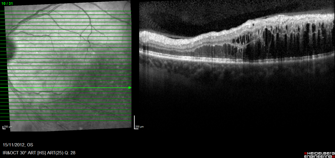
11
Advanced. Another pre-retinal haemorrhage showing a flat top.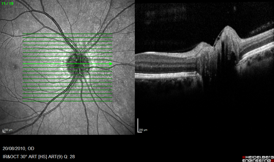
12
Advanced. This is an appearance very characteristic of a tractional retinal detachment where fibrovascular proliferation on the retinal surface extending into the vitreous has led to the retina being pulled away.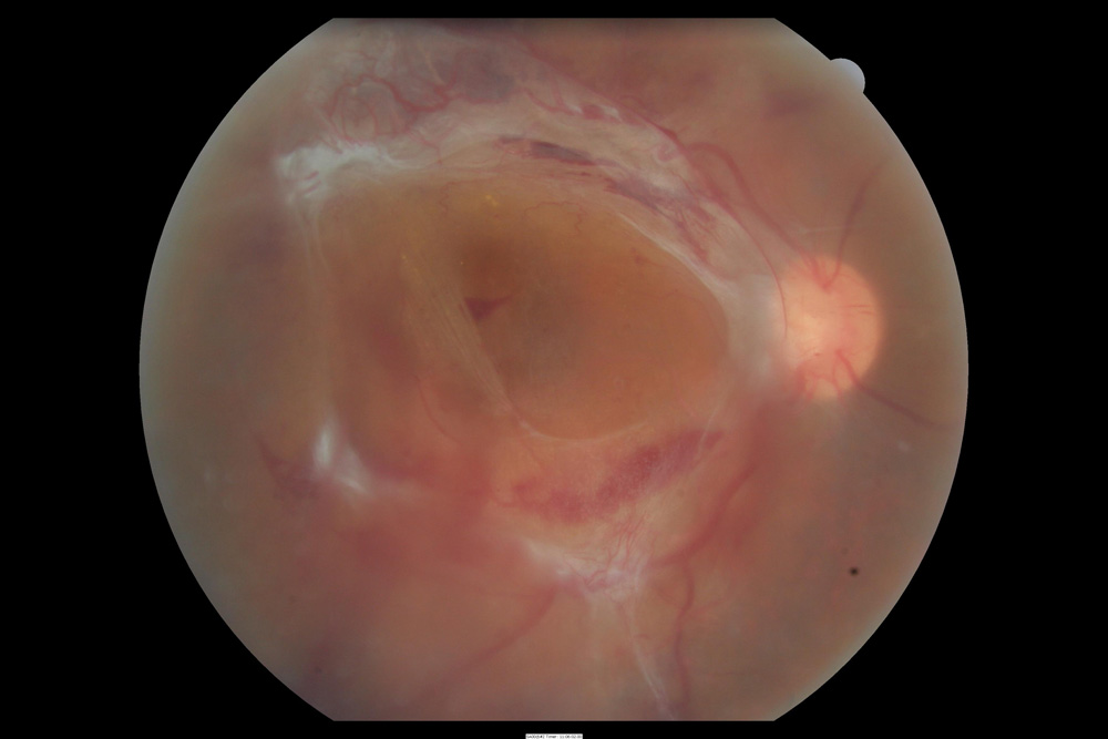
?
A comprehensive series on diabetic retinopathy will be published next year.