A CAUCASIAN MALE aged 36 years attended for an examination due to a rapid, constant reduction in vision in his left eye, first noted eight months ago and with no improvement to date.
At the time of the initial onset of reduced vision the patient went to his local Accident and Emergency department where he was diagnosed with a fungal infection (Candidiasis albicans).
The patient was an intravenous (heroin) drug addict and the possible cause of infection was by contaminated lemon/lime juice used in the preparation process of heroin or the use of non-sterile needles or syringes.
He was advised to be admitted into the hospital for a two-week course of antifungal therapy. The patient however, discharged himself and had not sought medical advice since. Visions were: 6/4-3 in the right eye and 6/60 in the left eye (no improvement with pin hole).
His last optometric examination was 10 years ago and he had not been prescribed spectacles or contact lenses. He was otherwise healthy, with no diabetes, glaucoma or high blood pressure noted. 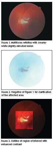
There were no symptoms of headaches, diplopia, flashing lights, pain, photophobia or floaters. The patient did report mild bilateral irritation/discomfort associated with poor lid hygiene and showed low-grade blepharitis. Refractive findings were negligible; right eye plano and left eye plano/-0.25 x 95. Intraocular pressures were within normal range; right eye 15mmHg and left eye 14mmHg (AO non-contact tonometry at 4:00pm).Ophthalmoscopy with dilated pupils and indirect ophthalmoscopy (Figure 1) revealed a multifocal retinitis with a creamy-white, slightly elevated lesion extending from the temporal side of the disc and two smaller, white, ‘snowballs’ a quarter of a disc diameter superiorly and one disc diameter temporally to disc within the macula region (to help visually clarify the affected areas see Figures 2 and 3).
The patient was advised to seek medical attention immediately and a letter was given to him to take to see his general practitioner that day for referral to the appropriate department for antifungal therapy. No glasses were prescribed and daily hot compresses and lid massaging advised to improve lid hygiene.
Candida albicans is a yeast-like fungus and is the most common fungal infection of the retina and choroid. They are commensal organisms that reside in the human body and are found normally in the female genital tract, the gastrointestinal tract, and the respiratory tract.
These fungi are usually kept in check by the host’s normal immune response.Factors that can predispose individuals to ocular candidiasis include intravenous drug use, long-term systemic use of antibiotics and cytotoxic agents, patients with long-term, in-dwelling catheters, prolonged hyperalimentation, corticosteroid therapy, recent abdominal surgery, alcoholism, poorly controlled diabetes, and patients with decreased immunity due to an underlying systemic disease (AIDS).
Patients are usually treated with intravenous amphotericin B or intravenous fluconazole and intravitreal injections of amphotericin B. Surgical treatment (therapeutic pars plana vitrectomy) is usually considered only in patients with dense vitreous opacities. Ocular symptoms can include visual loss, red eye, photophobia, pain, floaters, scotoma. The patient may be asymptomatic if the lesion is in the peripheral retina.
Register now to continue reading
Thank you for visiting Optician Online. Register now to access up to 10 news and opinion articles a month.
Register
Already have an account? Sign in here