We have had the Optos system at the City clinic for most of this year and it has proved a useful adjunct to ophthalmoscopy.
A good illustration of its application is shown by this patient case. Having been recommended to come to us for a 'thorough examination', this high myope was duly dilated and a thorough indirect ophthalmoscopic view undertaken.
It was immediately apparent that a whole swathe of inferior temporal retina was heavily pigmented, along with some peripapillary changes related to the patient's myopia.
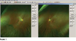
We then took images on the Optos system. Figure 1 shows the right and left maps displayed on screen simultaneously and clearly shows the area of pigmentation. The appearance suggests previous damage, such as an earlier detachment, but interestingly the patient reported no such incident.
Register now to continue reading
Thank you for visiting Optician Online. Register now to access up to 10 news and opinion articles a month.
Register
Already have an account? Sign in here