The Volk Pictor is a portable digital imaging device that has been around in UK optometry for some years now and has been taken up by practitioners requiring an imaging system able to operate in confined practice spaces and in domiciliary examinations. I first tried the Pictor some years back (Optician 11.11.11) and found it took a good deal of practice to achieve adequately clear images. Even then the images were of somewhat restricted field and the absence of any fixation point for the patient was a drawback. Furthermore, for a portable unit, the need for a USB feed for image transfer also prove a restriction.
The recently introduced Pictor Plus (UK distributor BIB Ophthalmic Instruments) keeps the best design features of its predecessor but has addressed the concerns. The unit is a lightweight handset (400g) onto which you attach the ‘retina module’ (300g). Assembly takes seconds and the overall weight should not bother any practitioner even when used for a prolonged period.
[CaptionComponent="2005"]An optional rubber cup (Figure 1) may be attached before approaching the patient (Figure 2) which serves as a buffer and guide as well as minimising the impact of external light.
[CaptionComponent="2006"]It is possible to capture retinal images through pupils as small as 3mm. An ‘anterior module’, weighing in at a meagre 92g (Figure 3) is also available for external and anterior imaging.
[CaptionComponent="2007"]There is now also available a slit-lamp mount (Figure 4) into which the unit in retinal capture mode slides allowing steady and micro adjustable positioning at the slit-lamp (Figures 5a and 5b).
[CaptionComponent="2008"][CaptionComponent="2009"][CaptionComponent="2010"]It would be better still if there was a version to allow the anterior module to be used at the slit-lamp – maybe something for the future?
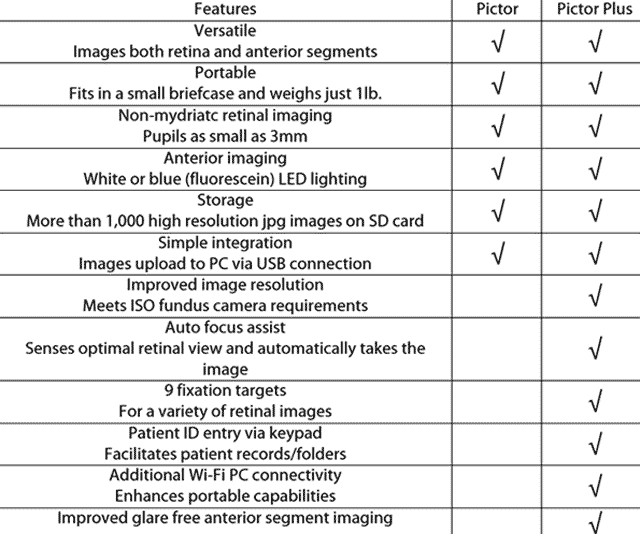
The camera offers 5 megapixel sensor resolution and images are stored on an internal SD card as Jpegs (up to 2,000 in any one session). The new unit can also capture and store video footage (in Mpeg-1 or Mpeg-4 formats). Table 1 offers a summary of the differences between the Pictor and Pictor Plus.
Improvements
The camera is programmed by a range of buttons around a colour LCD display screen. There is now the option of introducing a fixation target of variable brightness and this may be positioned in nine different locations to help centre an image on any particular retinal point. There is also the option to use a manual focus (for which you can introduce a spherical correction from +53.00DS to -78.00DS as well as an assisted autofocus or a full autofocus option.
Retinal photography is best done with the manual option. The light level may also be varied: increased for poor media patients for example, or decreased when getting too much reflection from the inner limiting membrane. When ready to go, you simply move towards the patient and (as the pupil is seen on screen) keep moving in, ensuring the pupil is centred until the retina hoves into view. When in focus, press the button and the image is stored in an instant. The first thing to notice is the wider field obtainable. Figure 6a was an image I took with the older Pictor.
[CaptionComponent="2011"]The new camera offers a 40-degree field (Figure 6b) with results not dissimilar to those from a desktop camera unit.
[CaptionComponent="2012"]Improvements in the image quality allow the image to be viewed on screen at up to six times magnification without loss of detail (Figure 6c).
[CaptionComponent="2013"]Capture can be pre-set to use white light (Figures 7, 8, and 9), white light and infra-red for a sharper view and red-free.
[CaptionComponent="2014"]All three are stored on the drive until transferred, or wiped at the end of a session. Patient data can be input on the new unit and images assigned accordingly.
[CaptionComponent="2015"]Better yet, like many high-end digital cameras, the unit has a Wi-Fi function allowing instant transfer to another database. I downloaded a free app on my tablet (Eye-Fi) and, once selecting the camera Wi-Fi in my settings, found that the images instantly appeared in my photos.
[CaptionComponent="2016"]This would be really useful when out and about without having to carry laptops and wires.
Anterior eye
For the anterior module, I found the autofocus more useful (Figure 10).
[CaptionComponent="2017"]The resolution was sufficient to allow a good degree of magnification again (for example, look at the lashes in Figure 11a at six times magnification – Figure 11b – or low grade blepharitis – Figure 11c).
[CaptionComponent="2018"][CaptionComponent="2019"][CaptionComponent="2020"]A blue light option can be set for angiography purposes but also proved capable of simple anterior fluorescein capture (Figures 12a and 12b).
[CaptionComponent="2021"][CaptionComponent="2022"]Welcome improvements
The Pictor Plus is a thoughtfully designed hand-held camera unit capable of significantly better image quality than its predecessor. I would suggest that the best images require some practice (though this also applies to desktop cameras too for undilated patients) but the expanded image options, wider field and ease of transfer make this a camera well worth using in a crowded practice, for taking out on domiciliary visits and for mobile screening.
Further information at http://volk.com/pictorplus and ophthalmic instrument site www.bibonline.co.uk

