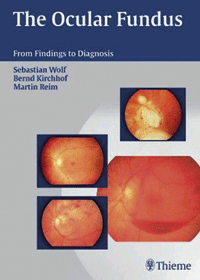
 Professor Robert Fletcher looks at a recently published text which successfully adopts an interesting and helpful format
Produced with 309 illustrations, including excellent coloured plates to Thieme’s high standard, much of this text is based on Aachen ophthalmic clinic material.
The layout is unusual yet very helpful; rather than a limited approach to fundus examination, a wide range of methods is presented and this should be particularly helpful to students.
Brief historical introductions concentrate on European classical and post-1950 techniques, embracing the bare bones of objective methods from photography, tomography, Doppler measurements, interferometry to angiography, as well as electrophysiological studies and ultrasound.
Thus, a broad introductory sweep can be enjoyed. References are somewhat biased towards continental papers and aspects of subjective assessments are somewhat curtailed.
Readers should not look askance at the presentation of visual field plots strictly in Goldmann modes – there is often an advantage in viewing isopter plots in this way, as in Figure 3.19, where sector defects of retinochoroiditis juxtapapillaris are shown.
There are suitable chapters in ‘appearances’ of fundus disorders, under different headings. Here are descriptions of an extensive variety of macular conditions, very well illustrated, backed by listed causes, comments on symptoms and indications of treatments. The section on areas of exudative retinopathy extends over nine pages and embraces one of the frequent tables, this one on risk factors for CRA occlusion.
Chapter 4 deals with the appearances of vascular disorders, emphasising the 78D lens and slit-lamp observation. Here the reader should appreciate the interplay between illustrations, concise and explanatory text and the emphasis provided in pageedge columns. The design is a pedagogic success.
Vitreous diseases are approached in Chapter 5, giving a good balance between symptoms and a variety of examination methods. Further chapters introduce optic nerve disorders, alongside physiological features and ‘disorders with no conspicuous fundus changes’, with several aspects given rather short shrift.
In addition to a very full index the authors have thoughtfully added several pages of systemic classification (with page numbers) and a list of abbreviations, a fitting finale to a most valuable book.
Register now to continue reading
Thank you for visiting Optician Online. Register now to access up to 10 news and opinion articles a month.
Register
Already have an account? Sign in here
