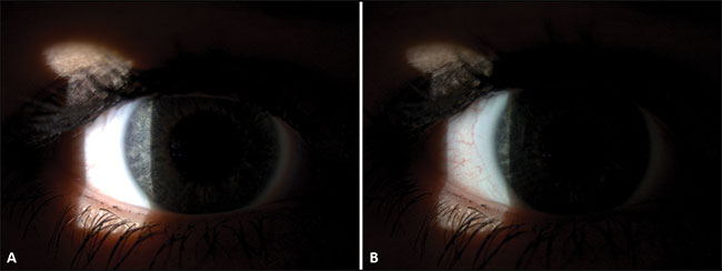
The slit-lamp biomicroscope is an essential part of the optometrists’ and contact lens opticians’ toolkit, enabling high magnification viewing of the anterior and posterior ocular structures.
When combined with an imaging system and the application of correct imaging techniques, the slit lamp can be used to capture high-quality photos and movies of the eye. These images can be useful for clinical documentation of ocular health, for patient education and, if necessary, as evidence in medicolegal situations.
This short series of articles will review some top tips that will help in producing high-quality slit-lamp photographs.
1 Reveal detail with the neutral density filter
The neutral density filter is a drastically underused part of the slit lamp. Without its use, finer detail can be missed. The neutral density filter modifies all wavelengths of light and avoids under and overexposure. In image A, the blood vessels of the conjunctiva cannot be seen due to overexposure; in image B, the addition of a neutral density filter enables the conjunctival blood vessels to be visualised.
2 Consciously Uncouple
Uncouple the beam from the lighting column (A) to enable retroillumination. This is an indirect illumination technique; the illumination does not fall directly on the structure being viewed. In retroillumination, the object of interest is illuminated from a light source behind it, with light being reflected off the retina to view the lens (B) or the iris to view the cornea. This technique creates a diffuse illumination, with any structure that does not transmit the reflected light appearing dark against a bright background. For example, lens opacities appear black against the reflected orange light.
3 Get the full picture
Use the diffuser to give a soft light, enabling capture of low magnification images of the anterior eye and adnexa. The diffuser scatters the light from the illumination system (B), enabling uniform illumination over the eye and creates a comfortable light for the patient (A without the diffuser versus C with the diffuser). When capturing images with the diffuser, ensure the slit-lamp beam width is completely opened and magnification is kept low (6-10x).
Read more
Part 2: Top tips for slit-lamp imaging
Part 3: Top tips for slit-lamp imaging
Dr Rachel Hiscox is clinical affairs specialist for Topcon GB