Features

Visual recognition in contact lenses. Part 1 - Silicone hydrogels

Lenses
Bill Harvey and David Ruston describe some clinical presentations related to silicone hydrogel lens fitting and follow up, indicating both their interpretation and how best to record them (C1471).

VISUAL RECOG1 F01Bill Harvey and David Ruston describe some clinical presentations related to silicone hydrogel lens fitting and follow up, indicating both their interpretation and how best to record them (C1471). 

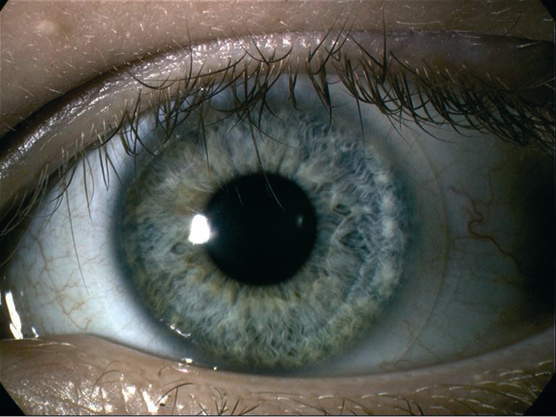
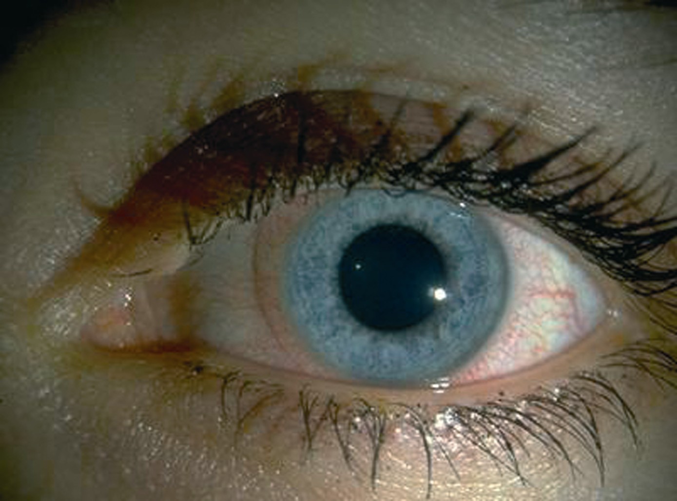
The increasing use of silicone hydrogel material contact lenses over recent years is related to a realisation of the excellent oxygen delivery properties of the lens resulting in a reduction in signs of hypoxia and serious keratitis. An immediate and impressive advantage is the apparent reduction in limbal and conjunctival hyperaemia seen with the material (Figure 1) as opposed to the more familiar hyperaemia seen with extensive wear of conventional hydrogels (Efron grade 1 trace hyperaemia in Figure 2 or grade 3 moderate hyperaemia in Figure 3).

PRE-FITTING ASSESSMENT
Many patients may be attracted by the publicity surrounding excellent oxygen properties or be recommended them by their practitioner. This is no more likely than when a patient has had previous problems of hypoxic stress. A classic sign of previous hypoxia is the presence of new vessels around the peripheral cornea (Figure 4).

It is important to establish firstly the likely cause of these. A localised pannus may be evidence of a response to an infective keratitis or severe inflammatory response, both of which might be contraindicative of contact lens wear, or at least extended use of such. Where the vascularisation is more generalised around the limbus, it is more likely to be a result of chronic hypoxia. The presence of ghost vessels (empty new vessels) is evidence of time having elapsed since the initial hypoxic insult. New vessels are often the most tangible evidence of the reason to move towards silicone hydrogels and though previous overwear of conventional hydrogels is not as prevalent as it used to be, there are still many patients that may present for silicone hydrogel fitting with new vessels present.

Measurement of the vessels is easily done by alignment of the slit over the vessels which may then be shortened to a height matching the vessel length. Efron has suggested that they should be measured from the point of visible iris and should not exceed 0.2mm for non-lens wearers or silicone hydrogel wearers, 0.4mm for daily wear RGP wear, 0.6mm for hydrogel daily wear and 1.4mm for extended wear of conventional hydrogels.1

Pre-assessment always should include an accurate corneal assessment including staining. Evidence of a diffuse superficial punctuate stain (Figure 5 shows moderate to severe corneal staining, Efron grade 3) may be a useful indication of inadequate tear flow or, rarely, some form of infective event. Diffuse stain may also be indicative of a toxic response, perhaps to a previous solution or some chemical agent introduced.

Figure 6 shows a classic 'smile stain' or inferior epithelial arcuate lesion. Reports of such lesions in conventional hydrogel wear are numerous.2-4 It is widely accepted that such a stain is evidence of either a metabolic influence or desiccation, such as might occur with poor tear flow under a previous lens, lens adherence or lens dehydration. Often a low grade smile stain may be best managed by refitting with a high transmissibility dehydration resistant material such as silicone hydrogel. A stain as significant as that shown in Figure 6 might benefit from a period of recovery without any lens wear before a new lens is fitted. The corneal compromise revealed by the stain may allow a point of entry for infection.

Blue or white light assessment may show evidence of previous infective activity. Deep stromal infiltrates may remain for many years after a severe adenoviral keratoconjunctivitis. Figure 7 shows a small isolated discrete scar from a previous bacterial infection. The nature of the bacterium might be deduced from the small size of the scar in this figure, gram positive infections generally causing smaller ulcers with less purulence than gram negative bacteria. Such evidence of an old infective keratitis is a significant finding, both in terms of a compromised cornea and also as an indication of a patient where microbial keratitis has been established. Good compliance and understanding must be sought in such a patient and a clear history with particular reference to the time elapsed since the infection was treated.

Evidence of meibomian gland dysfunction is an important finding in a pre-assessment of a potential contact lens patient. It has been estimated that up to 30 per cent of contact lens wearers show some loss of clarity of expressed meibomian secretion with around 11 per cent showing opaque oils.5 This is often seen in association with blepharitis and the possibility of the Staphylococcus epidermis flora around the lid margins leading to complications if lenses are to be fitted for continuous or extended wear purposes. However, gland dysfunction should be considered as a distinct entity from posterior blepharitis and the opaque secretion may lead to greasing of the contact lenses and dry eye symptoms. Figure 8 shows inspissated secretion from the meibomian glands. Expulsion of the material (Figure 9) shows typical debris and opaque material in the tear film (Figure 10). Expression of debris may be useful in management if done carefully alongside lid scrubs and warm compresses, the latter again aiding the expression of the abnormal secretion. Alhough these measures help control the condition, its chronic nature makes it a recurrent nuisance.

Figure 11 shows meibomian gland involvement alongside both anterior and posterior blepharitis signs. There is clearly redness and irregularity of the lid margin, blockage of the glands, and the familiar crusting and matting of the lashes. Such an appearance would need treating before any continuous lens modality was considered. Severe anterior blepharitis is evident and common without meibomian involvement, as shown in Figure 12. Again lid cleaning on a regular basis is advisable.

FITTING CHARACTERISTICS
Silicone hydrogels are available with a maximum of two base curves, so the lenses either fit or do not with little leeway for modification. In the occasional instance where a lens is too flat, the sometimes very high modulus results in the rucking up of the lens edge (or fluting as it is known) as shown in Figures 13, 14 and 15 (courtesy of Brian Tompkins). Such an appearance demands a refit with a steeper lens or an alternative brand of lens of lower modulus.

POSSIBLE COMPLICATIONS OF WEAR
Most of the complications resulting from the extended or continuous wear of silicone hydrogels have now been well documented. Superior epithelial arcuate lesions were thought of as a rare complication until the rise in numbers wearing the much stiffer silicone hydrogels from the late nineties onwards. As shown in Figure 16, the lesion presents as a characteristic arcuate ('inverted smile') epithelial lesion typically 2 to 3 mm within the cornea from around 10 to 2 o'clock. Sometimes symptomatic, the lesion still represents a compromise of the epithelium and a move to a lower modulus material is usually indicated.

Figure 17, as well as showing significant hyperaemia, also merits careful study as at around 4 o'clock a small area of localised infiltration is visible at around 1mm within the cornea. This isolated peripheral size and location is indicative of a contact lens induced peripheral ulcer or CLPU. This, together with the contact lens induced acute red eye or CLARE (marked in Figure 18 and mild in Figure 19) were classic of what was until recently described as sterile infiltrative keratitis as distinct from infective microbial keratitis. The latter is characterised by pain, significant staining, anterior chamber activity, central location and possible discharge, some of which are seen in Figure 23. The former could be managed within practice, usually with a period of non-lens wear, while the latter needs immediate referral to an ophthalmologist for anti-infective treatment. Recent research has, however, blurred this dichotomy somewhat.6 It has now been suggested that keratitis is instead best thought of as a spectrum of severity. Continuous wear increases the likelihood of severe keratitis, but with silicone hydrogels this is more likely to be the less serious type of presentation while with conventional hydrogels a serious infective keratitis is more likely (five times in fact in one study6).

A high modulus lens and a prolonged mechanical action on the palpebral conjunctiva is associated with an increased incidence of contact lens induced papillary conjunctivitis. Use of fluorescein (Figure 20a) helps heighten the observation of papillae on the palpebral conjunctiva. In some cases, the papillary changes can be very localised. this is particularly the case where there has been edge fluting (Figure 20b). In these cases, removal of the allergenic factor (contact lens wear) is essential and therapy may include anti-histamines, mast cell stabilisers and the use of topical lubricants to aid comfort. Rarely is it necessary for anti-inflammatories to be used.

Such complications, albeit all manageable, are obvious reasons why regular aftercares with fluorescein assessment are essential with extended wearers of contact lenses. Figure 21 shows such an assessment after five weeks of continuous wear. Small epithelial staining clusters and a rapid break up time are clearly seen. Figure 22 shows a more diffuse inferior epithelial punctuate stain possible indicative of a significant tear flow compromise.

Perhaps the best remembered but possibly least clinically significant complication of high modulus lenses is the presence of mucin balls (Figure 24). Staining may simply reflect pooling within epithelial depressions made by the mucin clumps but does not indicate epithelial compromise.

PATIENT SATISFACTION
It is perhaps obvious that comfort and satisfaction are the ultimate requirements of practitioners and patients alike, but these cannot be captured visually, unlike complications. However, the material properties that may lead to comfort can be listed as:

  • Lens design
  • Oxygen delivery
  • Wettability
  • Lubricity
  • Modulus of elasticity.

    The challenge for manufacturers is to produce silicone hydrogels that have the appropriate balance in these properties to deliver real benefits to wearers. Second generation silicone hydrogels are now coming to the market that claim to address these issues. Adequate oxygen delivery to the cornea is now assured by all of the available products. The issue for patients and practitioners is now to identify those products that really deliver comfort benefits for patients.
    The modern world throws up numerous challenges for practitioners and patients alike. These include air-conditioned offices, extended computer use, smoky polluted environments, frequent flying and additionally the use of medications such as anti-histamines and beta-blockers that lead to ocular dryness. The success of these second generation lenses will perhaps depend on their ability to meet these challenges and deliver on those attributes that patients are seeking to ensure long-term comfortable contact lens wear.

    LEARNING OBJECTIVES
    The main clinical appearances to look for before, during and after fitting silicone hydrogel lenses have been described and illustrated.

  • ACKNOWLEDGEMENT
    Figures 13-15 are courtesy of Brian Tompkins.

    References
    1 Efron N. Contact Lens Complications. P157. Butterworth-Heinemann (2005)
    2 Zadnik K, Mutti DO. Inferior arcuate staining in soft contact lens wearers. International Contact Lens Clinic, 1985; 12, pp110-113.
    3 Little SA, Bruce AS. (1995). Role of the post-lens tear film in the mechanism of inferior arcuate staining with ultrathin hydrogel lenses. CLAO, 21, pp175-181.
    4 Watanabe K, Hamano H (1997). The typical pattern of superficial punctuate keratopathy in wearers of extended wear disposable contact lenses. CLAO 23, pp134-137.
    5 Ong BL, Larke JR. Meibomian gland dysfunction: some clinical, biochemical and physical observations. Ophthalmic and physiological optics, 1990; 10, pp144-148.
    6 Morgan PB, Efron N, Hill EA, Raynor MK, Whiting MA and Tullo AB.  Incidence of keratitis of varying severity among contact lens wearers. British Journal of Ophthalmology, 2005;89:430-4.

  • Bill Harvey is optician clinical editor. David Ruston is director professional affairs UK and Ireland, Johnson & Johnson Vision Care