A seven-year-old girl slips in the bathroom, and breaks her leg (fracture in femur). A six-year-old boy while playing, jumps from a stool, and fractures his leg. A 52-year-old businessman falls down from the upper berth while travelling in a train, but is unhurt. And, a 65-year-old housewife falls from her bed while sleeping, and is apparently unhurt; let us call her Mrs Grewal.
Sensing something wrong with her progressive-power spectacles, Mrs Grewal goes for an eye examination. She is a hypermetropic astigmatic presbyope, and is not satisfied with improvement in vision with glasses in her right eye.
In this article I will first consider the nature of macular holes. I will then review their assessment and management. Then we will return to the case of Mrs Grewal.
What is a macular hole?
A macular hole is a small break in the macula, located in the centre of the retina. The macula provides sharp, central vision, and a macular hole can cause blurred and distorted central vision (figure 1). Macular holes are related to aging and usually occur in people over the age of 60. Macular holes are different than age-related macular degeneration, although symptoms in both the conditions are similar and both are common in people 60 and over.1
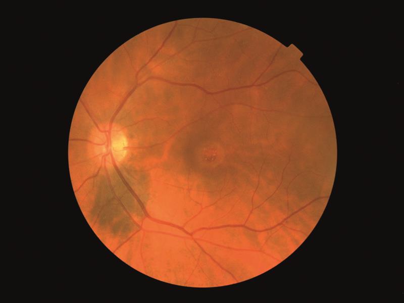
Figure 1: Fundus photo of a full thickness macular hole with drusen-like deposits
Aetiology
The posterior chamber of the eye (about 80% of the eye) is filled with vitreous gel that helps the eye maintain its round shape. Vitreous contains innumerable fine fibres that are attached to the surface of the retina. Ageing slowly pulls away vitreous from the retinal surface. The area where vitreous contracts, gets filled with natural fluids. The process is normal, and mostly there are no adverse effects, except in some people who may experience a small increase in floaters that seem to swim before the eye. If, however, vitreous is firmly attached to the retina when it pulls away, it can tear the retina and create a macular hole. Otherwise, when vitreous pulls away from the retina, some of the remaining fibres on the retina may contract, increasing tension on the retina, leading to a macular hole. In both cases, the replaced natural fluid can seep through the hole onto the macula, blurring and distorting central vision. Macular hole can also occur in high myopia, due to trauma to the eye, or in those having retinal detachment, and risk factors include diabetes, history of retinal tear or detachment, and inflammation in the eye (uveitis).2
Symptom
The most common symptom of a macular hole is a gradual decline in central vision of the affected eye, that can occur as blurring, distortion, or a dark spot in central vision.3
Clinical Assessment and Diagnosis
Important elements of the patient history include the duration of symptoms (as this influences prognosis), any significant preceding ocular trauma, and the existence of high myopia. A history of retinal vascular disease and ocular inflammation should be sought.
Visual impairment in stage 1 and 2 macular holes is usually mild-to-moderate (6/7.5 – 6/18), but progresses to around 6/30 – 6/120 in stage 3 and 4. In general, any diagnosis of full thickness macular hole (FTMH) in an eye with visual acuity of 6/12 or better should arouse suspicion.
A yellow spot or ring (stage 1) or full thickness foveal defect (stages 2-4) is usually evident on careful ophthalmoscopy. A cuff of subretinal fluid, intraretinal oedema, and drusen-like deposits may also be seen (figure 1). Long-standing cases may have retinal pigment epithelial changes at the margin of the subretinal fluid. Accompanying epiretinal membranes are not uncommon.
The Watzke-Allen test involves projecting a narrow beam of light across the macular hole using a slit lamp, and is considered positive if the patient reports seeing a gap in the light. Although a positive test is consistent with the presence of an FTMH, research since the advent of OCT imaging has shown that this test has a sensitivity of only 60%. As such, it is now largely redundant but may be helpful if the view is suboptimal or you do not have access to OCT.
OCT imaging is used to document the presence of a hole, allow accurate staging, eliminate differential diagnoses, and educate the patient about their condition. The foveal dehiscence is usually obvious, and retinal oedema at the margins with a cuff of subretinal fluid is usually seen.
Optical coherence tomography (OCT) is the current gold standard in the diagnosis, staging, and management of macular holes (figure 2). This quick, non-invasive imaging technique allows for evaluation of the macula in high resolution using reflected light, and helps differentiate a hole from other eye conditions with similar symptoms. No laboratory tests are needed in cases of idiopathic macular holes (those without a known cause).4

Figure 2: Single line scan OCT through a full thickness macular hole
Staging
The staging system for an FTMH was first proposed by Gass and later revised. Stage 1 refers to subtle macular changes that precede the development of a true full thickness hole – stage 1A (impending hole) is seen as a yellow spot at the fovea, and 1B (occult hole) as a tiny yellow ring. Stage 2 refers to a full thickness macular hole less than 400 microns in diameter, incompletely bridged with posterior hyaloid (vitreous cortex). Stage 3 describes an FTMH greater than 400 microns in diameter, with localised posterior hyaloid separation from the macula. A pseudo-operculum may be seen suspended on the posterior hyaloid overlying the fovea. And Stage 4 refers to an FTMH with complete posterior vitreous detachment. Staging is important because it has prognostic implications which are discussed below. Macular holes tend to progress through these stages over weeks to months with worsening vision and metamorphopsia.
The characteristics of the different grades of macular hole are summarised in table 1.

Table 1: Characteristics of macular holes graded stage 1 to 4
The hole tends to gradually enlarge and the central vision gradually worsen before stabilising at approximately 6/60 to 6/120. Peripheral vision is generally unaffected; however retinal detachment occasionally develops (particularly in high myopes) which may compromise peripheral vision even with anatomically successfully repair. The prognosis for established macular holes (stages 2-4) is poor without treatment, as spontaneous closure is uncommon (approximately 10% of stage 2 holes, less than 3% of stage 3 holes, and very rarely with stage 4 holes).
The risk to the fellow eye is also substantial, with approximately 15% developing a full thickness macular hole within five years. However, the risk to the fellow eye is understandably much lower (approximately 2%) if it has already had a posterior vitreous detachment, as this is the event that typically causes the hole in predisposed patients.
Management
Although some macular holes can seal themselves and require no treatment, surgery is invariably necessary to help improve vision.

In this surgical procedure – called a vitrectomy – the vitreous gel is removed to prevent it from pulling on the retina and replaced with a bubble containing a mixture of air and gas. The bubble acts as an internal, temporary bandage that holds the edge of the macular hole in place as it heals. Surgery is performed under local anesthesia and often on an out-patient basis (and sometimes by admitting the patient for one day). Following surgery, patients must remain in a face-down position, normally for one week but sometimes for up to three weeks. This position allows the bubble to press against the macula and be gradually reabsorbed by the eye, sealing the hole. As the bubble is reabsorbed, the vitreous cavity refills with natural eye fluids. Maintaining a prone position (maybe, by way of the specially-made pillow – figure 3, pictured left) is crucial to the success of the surgery.
The most common risk following macular hole surgery is an increase in the rate of cataract development. In most patients, a cataract can progress rapidly, and often becomes severe enough to require removal, and, therefore, many surgeons do a combined cataract and vitrectomy procedure. For a few months after surgery, patients are not permitted to travel by air, as changes in air pressure may cause the bubble in the eye to expand, increasing pressure inside the eye. Vision improvement varies from patient to patient. People that have had a macular hole for less than six months have a better chance of recovering vision than those who have had one for a longer period. Vision recovery can continue for as long as three months after surgery.5
The case in point
Coming back to Mrs Grewal’s case, she is not satisfied with 6/9 vision in distance and N8 vision in near with glasses in her right eye, and insists with more concern that she misses a letter or two on distance as well as near test types; she also sees objects slightly distorted. She has earlier been told about a minor lenticular opacity that was not likely to increase in near future. IOP (intraocular pressure) measurement and direct and indirect ophthalmoscopy indicate VFA (visual field analysis), but she is told there was no urgency.

Figure 4: Pre-operative appearance of macular hole
She returns to the clinic after about four months with the complaint that she now notices (with her right eye) a break in the tube-light. Urgent VFA and OCT tests are now advised. The OCT test is done first, and it confirms the presence of a macular hole, that is reconfirmed by way of indirect ophthalmoscopy, and Mrs Grewal is advised immediate combined cataract and vitrectomy surgery, to which she agrees (though with an element of indecisiveness and apprehension)…and now, after four months, having recuperated seems to be feeling much better (except minor irritation that can at best be attributed to individual feeling). Figures 4 and 5 present pre- and post-operative OCT pictures respectively.

Figure 5: Post-operative appearance of macular hole
In conclusion
Macular holes can cause a significant drop in central vision. In many cases vision is restored, either through natural resolution or after surgical intervention. The nature of the hole is assessed and can be graded as an indicator of the likelihood of success of treatment. There are a variety of causes of macular hole, including high myopia, retinal detachment, diabetes, chorioretinitis and trauma. This paper highlights one case where what might be considered a minor traumatic incident appears to have instigated macular hole formation and underlines the importance of careful questioning alongside fundus examination (including with OCT) where anyone reports sudden or recent central acuity changes.
Dr Narendra Kumar holds a diploma in optometry (DROpt), degree in ayurvedic medicine and surgery (BAMS), and certificate in rehabilitation (PGCR) qualifications and is a member of IACLE. Having served as refractionist at Sir Ganga Ram Hospital for 30 years, he now looks after contact lens cases at his ophthalmologist son’s clinic OphthaCare Eye Centre. He has edited the journal Optometry Today since 1970, is the chairman of the charitable trust Eye Care India, and the author of Ophthalmic Dispensing Optics and Babloo goes for an eye test books and ‘I care for eye-care’ bookmark.
References
1 National Eye Institute, http://nei.gov/health/macularhole.
2 Dr Charu Gupta, Retinal Surgeon, Ophthacare Eye Centre, Janakpuri, New Delhi, ophthacare@gmail.com.
3 Dr Maneesh Kumar, Oculoplasty and Phaco Surgeon, Ophthacare Eye Centre, Janakpuri, New Delhi, ophthacare@gmail.com.
4 American Society of Retina Specialists, www.asrs.org.
5 Dr Noshir Shroff and Dr Cyrus Shroff, Shroff Eye Centre, New Delhi, www.shroffeye.org.
Useful reading
Morten la Cour and Jakob Friis. Macular holes: classification, epidemiology, natural history and treatment. Acta Ophthalmol Scand. 2002: 80: 579-587.