The term anisometropia is used to describe the clinical situation that exists when the dioptric values of a patient’s right and left spectacle corrections are unequal and can be described as low (0.25D to 0.75D), medium (1.00D to 2.00D) or high (>2.25D). The term antimetropia is sometimes used if one eye is myopic and the other hypermetropic.
Anisometropia can give rise to two optical problems. Aniseikonia, meaning not equal images, refers to the difference in cortical image sizes and has the potential to disturb binocular vision. It occurs as a result of unequal spectacle magnifications due to the back vertex power, form, thickness and vertex distance of the correcting spectacle lenses. Secondly, when the eyes of anisometropic subjects rotate to view through points away from the optical centres of a pair of spectacle lenses, different prismatic effects between the two eyes may be experienced. This difference in prismatic effects is known as differential or relative prism.
Due to the existence of large horizontal fusional reserves and smaller horizontal eye movements when reading, differential prismatic effects in the horizontal are rarely a problem. However, if differential prismatic effect occurs in the vertical meridian, it can create problems by again disturbing binocular vision.
An additional consideration in anisometropia is the mechanical appearance of the two lenses as one lens will often be thicker and heavier than the other. It is possible to use combinations of lower and higher refractive index materials and/or aspheric and non-aspheric lens forms in order to try to balance the weight and appearance of the right and left lenses. However, care must be taken if reflection-free coatings are used as the residual reflection may not be equal if different refractive index materials are used. Parts 17 and 18 of this series will consider differential prismatic effects in more detail.
Spectacle magnification
When a spectacle (or contact) lens correction is worn, the retinal image formed is referred to as the corrected retinal image. With reference to the eye when uncorrected, the image formed is referred to as the basic or uncorrected retinal image. As far as corrected retinal image sizes are concerned; positive spectacle lenses produce an increase when compared to the basic retinal image size, while negative lenses produce a decrease. It follows that positive lenses make things appear larger to the subject, and negative lenses have the opposite effect. These effects are exaggerated by increasing the vertex distance and changing the form of the lens. Spectacle magnification is the term used to describe the ratio of the size of the retinal images formed in the same eye when corrected and uncorrected. Spectacle magnification is the magnification of the retinal image produced by placing a correcting lens in front of an eye and is defined as the retinal image size in the corrected ametropia eye over the retinal image
size in the same uncorrected ametropia eye, or:
As spectacle magnification is a ratio it has no units. The simplest way to calculate the size of the retinal image formed when the eye is corrected is to calculate the spectacle magnification and multiply it by the uncorrected (basic) retinal image size. The size of the corrected retinal image is therefore given by:

Spectacle magnification with thin lenses
Any spectacle magnification produced by a ‘thin’ lens is due to the power and position of the correcting lens only as a thin lens has no form or thickness. The magnification produced by a thin spectacle lens is known as the power factor (PF). For thin spectacle lenses, spectacle magnification (or power factor) can be given by:
This definition and equation applies to hypermetropia and myopia, both axial and refractive. An alternative expression to find the spectacle magnification or power factor of a thin lens is:
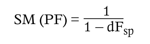
where d is the vertex distance in metres and Fsp is the power of the thin lens in dioptres. The above expression assumes that the entrance pupil of the eye coincides with the reduced surface. If the position of the entrance pupil is taken into account it is usually assumed to be 3mm to the right of the corneal surface. The symbol a is used in place of d and a = d + 3mm. So,

Thick spectacle lenses
Any spectacle magnification produced by a thick or ‘real’ spectacle lens is due to the power and position of the lens and also the form and thickness of the lens. The magnification due to the form and thickness of a lens is known as the shape factor (SF) which is given by:
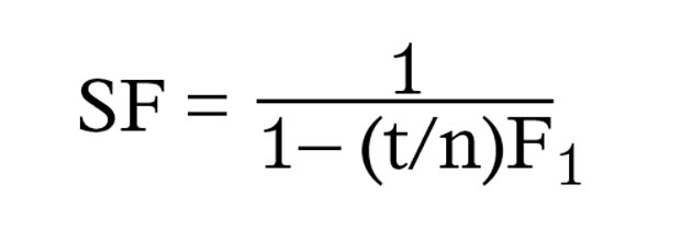
where t is the centre thickness of the spectacle lens in metres, n is the refractive index of the lens material and F1 is the power of the front surface of the lens in dioptres. An alternate expression to find the shape factor of a thin lens is:
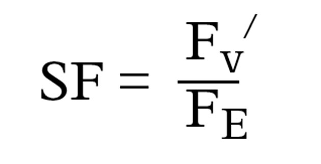
where Fv/ is the back vertex power of the thick lens and FE is the power of the equivalent thin lens. This alternative expression is mainly used in contact lens problems involving gas permeable (RGP) contact lenses.
So, unlike a thin lens, the magnification produced by a thick spectacle lens has two components, the power factor and the shape factor. The total spectacle magnification for a thick lens is therefore:
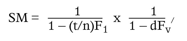
Fv/ is the back vertex power of the thick lens.
Shape factor is important for plus lenses and cannot be ignored. However, it can be ignored for minus lenses. Why? For a minus lens, the centre thickness will be in the region of 1.5mm. If we take the refractive index to be 1.5, t/n will be in the region of 1.0mm. This means that the shape factor value will always be in the region of 1.0. Put some numbers into the expression and try it! If the spectacle magnification for a thick lens is PF x SF, and if the SF = 1.0, then PF x 1.0 = PF. The shape factor can therefore be ignored for a minus lens. For plus lenses, the shape factor always has a significant effect on the SM produced and cannot be ignored.
How much spectacle magnification can be noticed? Researchers have found that a difference of as little as 0.25 per cent in the spectacle magnifications between the right and left eyes of a patient affected a subject’s binocular vision function. On a more practical note, a patient will notice the magnification produced by a pair of +1.00D reading lenses. As a rule of thumb, each 1.00D of lens power gives approximately 1 per cent magnification. The patient is therefore noticing 1 per cent spectacle magnification. Table 1 gives typical values for the spectacle magnification (distance vision) of a range of lenses. In each case, when calculating the power factor, the position of the entrance pupil of the eye (a) was taken to be 16mm from the back vertex of the correcting lens.
Spectacle magnification can also be expressed as a percentage. For example a value of 1.188 is equivalent to a 18.8 per cent ((1.188 – 1.000) x 100) increase in image size whereas a value of 0.942 is equivalent to a 5.8 per cent ((1.000 – 0.942) x 100) decrease in image size.
Some practical effects of spectacle magnification
When dispensing a first-time prescription, for example -2.50DS R & L, the patient should be forewarned that the image will be smaller than without glasses. Sometimes, when not warned, such first-time wearers do not like the smaller image, even though it is clearer.

A difference in the spectacle magnification between the right and left lenses can cause problems with binocular vision. However, such problems only occur if the difference in the right and left spectacle magnifications is greater than about 5 per cent. Binocular vision with spectacles in the case of monocular aphakia with the other eye being normal is the most dramatic example of this. In this situation, the difference in spectacle magnifications can be as great as 30 per cent. Problems are caused because the induced aniseikonia (a difference in cortical image sizes) is such that images formed in the two eyes do not fall on corresponding areas. Thus cortical cells are not driven binocularly.
In astigmatism, correction with spectacles produces different magnifications in the two principal meridians. This can give rise to perceived distortion of space, which can be made worse if the front surface of the lens is toroidal in a plus prescription because the shape factor compounds the problem.
When changing from spectacles to contact lenses, the retinal image size is larger in myopia but smaller in hypermetropia. The opposite is true when going from contact lenses to spectacles. This is particularly noticeable with higher myopic prescriptions, for example, a -10.00D myope may have a visual acuity of 6/9 with spectacles and 6/6 with contact lenses.
Changing the form of the lens will change the spectacle magnification and therefore the retinal image size especially with plus lenses as they are thicker than minus lenses. In anisometropia (a difference in the right and left prescriptions) where a patient is adapted to particular right and left lens forms, it is important to maintain those forms so that binocular vision remains comfortable in the new spectacles. This could be important when changing from spheric forms to aspheric forms.
Patients prescribed with reading spectacles for the first time think that they are just for ‘magnification’. The magnification with a pair of +1.00DS spectacle lenses is about 1 per cent which, although perceptible, is of no practical value when compared with magnifications of 2X (ie 100 per cent) or more which can be obtained with magnifiers.
One of the advantages of aspheric lenses (especially plus aspheric lenses) is that they exhibit less spectacle magnification. This is because the lens is flatter than the spherical equivalent and therefore produces less shape factor magnification. It should however be noted that some patients may dislike the smaller retinal image size produced as a consequence of a reduction in shape factor magnification. Aspheric lenses can therefore be problematic when prescribed for near vision if the patient has age-related macular changes.
Aniseikonia
The most impressive, and these days rare form of anisometropia encountered in practice is probably unilateral aphakia. In this situation, the crystalline lens has been removed from one eye and has not been replaced with an intraocular lens. If the aphakic eye is corrected using a spectacle lens the retinal image will be magnified to such an extent (˜ 30 per cent) that binocular vision may not be possible as fusion of the retinal images may only occur in a very small region close to the foveas.
As far as phakic subjects are concerned, completely normal binocular function is not often found in patients with more than 5 per cent aniseikonia when wearing a spectacle or contact lens correction. In practice the rule of thumb ‘1 dioptre of power produces 1 per cent spectacle magnification’ is often used to estimate the aniseikonia present although this really only applies to ‘thin’ lenses.
Symptoms/signs of aniseikonia in corrected subjects may include suppression of the image in one eye, poor stereopsis and ocular discomfort/headaches. A change from spectacles to contact lenses (or contact lenses to spectacles) when a subject has anisometropia may induce binocular vision problems due to aniseikonia. Where an anisometropic patient is adapted to spectacles, it is important to maintain the form of the lenses in any new spectacles since changing the form will relatively affect the retinal image sizes and therefore the aniseikonia. This is particularly important in hypermetropic cases where the lens thickness cannot be ignored, as it can in myopic spectacle corrections.
Very occasionally spectacle lenses are used to alter the size or shape of the retinal image without changing its position. Such lenses are known as iseikonic lenses and are used to equalise the right and left spectacle magnifications and therefore size of the right and left retinal images. Iseikonic lenses can on rare occasions be used in cases of symptomatic aniseikonia resulting from anisometropia. However, aniseikonia can also be induced by a pair of lenses of equal power but different form and thicknesses. Iseikonic lenses usually contain a refractive power but they can be plano. They are also known as size lenses as they change the size but not the position of the retinal image. When designing an iseikonic lens the power factor is determined by the prescription and vertex distance. The lens designer can therefore alter the shape factor to produce the required spectacle magnification as increasing the curvature of the lens or increasing its thickness produces an increase in magnification. It is usual to increase the magnification of the weaker of the two lenses. A standard lens is used for the higher plus (weaker minus lens) of the pair. A pair of lenses of unequal back vertex power but equal spectacle magnifications is called an isogonal pair.
Spectacle magnification and contact lenses
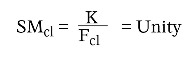
When a hydrogel contact lens is fitted to an eye, the lens ‘drapes’ over the eye and follows the curvature of the anterior ocular surface. This implies that the tear lens formed between the contact lens and the cornea (if indeed there is one) will have zero power, and the ametropia is corrected by the back vertex power (BVP) of the hydrogel contact lens. As the vertex distance of a contact lens is zero, we can in most cases assume that in order to correct a patient’s ametropia, the BVP of a hydrogel contact lens will be close to the patient’s ocular refraction (K). In other words FCL = K. If the power of the correcting spectacle lens along with its vertex distance is known, K can be calculated using the expression:
where d is the vertex distance in metres.
However, when an RGP contact lens is placed on the eye, the back surface of the contact lens maintains its shape and a tear lens of predictable form and power is formed between the rigid contact lens and the cornea (Figure 1). The patient’s ametropia is, therefore, corrected with a contact lens/tear lens system and the BVP of the RGP contact lens will not (unless an afocal tear lens is formed) be the same as the patient’s ocular refraction.
[CaptionComponent="2764"]
The contact lens/tear lens system formed when a RGP contact lens is placed on an eye means that three elements are involved in the formation of the final retinal image: the contact lens, tear lens and the eye. The vergence that actually corrects the patient’s refractive error is the vergence leaving the back surface of the tear lens L4/ (Figure 2).
[CaptionComponent="2765"]
When performing a paraxial ray-trace through a contact lens/tear lens system, it is often assumed that a thin air gap exists between each element, which can simplify the calculation of surface powers and vergences (Figures 1 and 2). The tear lens formed by an RGP contact lens is shown in Figure 3.
[CaptionComponent="2766"]
In practice, myopic patients are often told that they may obtain slightly better distance vision with their contact lenses than with their spectacles. Hypermetropic patients may be told the opposite. These potential differences in acuity are caused by differences in spectacle magnification when comparing correction with spectacles and contact lenses.
For thin lens systems and model eyes, where the entrance pupil coincides with the cornea or the reduced surface, spectacle magnification is given by:
When considering hydrogel contact lenses, as the tear lens is usually considered to be plano and the contact lens thin, we can assume that the power of a hydrogel contact lens is equal to the eye’s ocular refraction. The spectacle magnification produced by the hydrogel contact lens is therefore:
When considering contact lens systems, the entrance pupil of the eye is normally taken to be 3mm behind the corneal surface and both the contact lens and the tear lens have a thickness. In such cases, we are not therefore able to simply state that the spectacle magnification provided by a contact lens system is equal to unity.
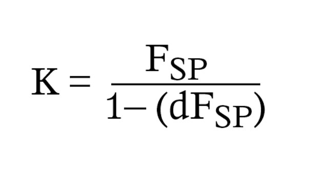
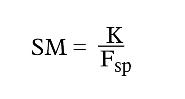
For a contact lens tear/lens system, the spectacle magnification produced is the product of the power factor and the shape factor, the expression for which is:
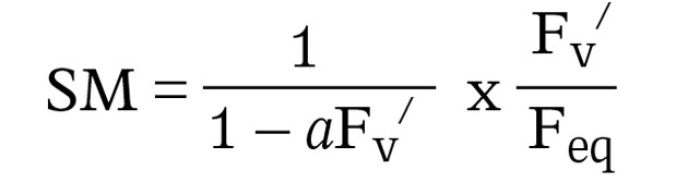
where:
- Fv/ is the BVP of the contact lens-tear lens system (L4/ in Figure 2)
- Feq is the equivalent power of the lens system
- a is the distance from the back vertex of the lens system to the entrance pupil of the eye.
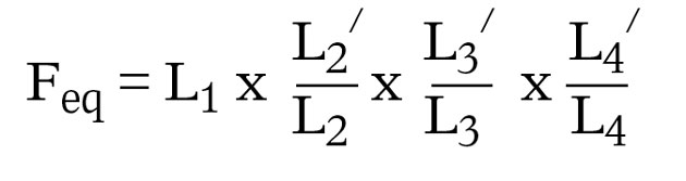
For spectacles a = vertex distance + 3mm and for contact lenses a = 3mm as d = 0. For a contact lens tear/lens system, the equivalent power is given by:
The above vergences are shown in Figure 2.
As an example, a patient is corrected using a spectacle lens of power +10.50DS at a vertex distance of 12mm. The back surface power is -2.00D, centre thickness 6mm and refractive index 1.60. The spectacle magnification produced by this correction would be in the region of 25 per cent. If the same patient is fitted with a RGP contact lens, where the contact lens centre thickness is 0.3mm and the tear lens thickness is 0.1mm, the spectacle magnification produced would be approximately 5 per cent. Even though the thicknesses of both the RGP contact lens and the tear lens are small compared to the thickness of the spectacle lens, the steep curves involved mean that the spectacle magnification produced is significant.
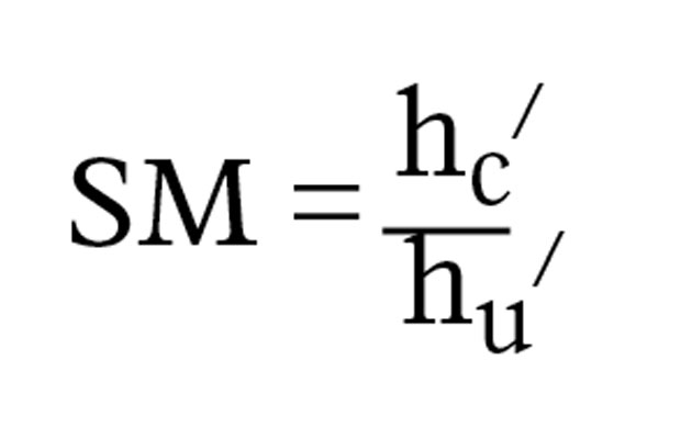
The most commonly encountered refractive error where there are marked orthoptic advantages to wearing contact lenses is anisometropia. When considering aniseikonia in both spectacle and contact lens correction, it is necessary to calculate the relative spectacle magnification produced. Relative spectacle magnification for a given distant object is defined as the retinal image size in the corrected ametropic eye (hc/) over the retinal image size in the standard emmetropic eye (hem/):
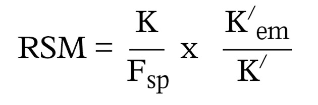
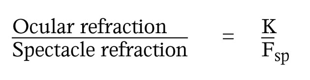
In the standard emmetropic eye, K/em will always be equal to +60.00D, so
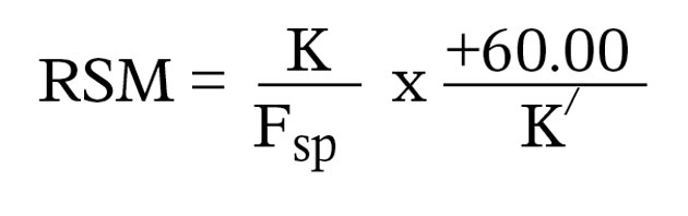
where K/ is the dioptric length of the ametropic eye (K/ = K + Fe), K is the ocular refraction and Fsp is the spectacle refraction. The significance of relative spectacle magnification is the comparison of corrected retinal image sizes in myopic and hypermetropic eyes with the basic retinal image size in a standard emmetropic eye. It is also interesting to consider any differences in aniseikonia if the patient’s refractive error is axial or refractive in origin. In axial ametropia, spectacles are theoretically better as any aniseikonia will be less and binocular vision will be more comfortable. The opposite is true if the anisometropia is refractive in origin, as correction with contact lenses will result in the right and left retinal images being the same size. However, this theoretical prediction, known as Knapp’s law, was disproved by research that revealed that contact lenses reduce aniseikonia in all forms of anisometropia.2 In addition, refractive (non-strabismic) anisometropes are likely to achieve their best binocular visual acuity and stereoacuity when wearing contact lenses as opposed to spectacles.3
Refractive correction without patching can improve the best-corrected acuity in an amblyopic eye and this therapeutic effect may be enhanced with contact lenses. It is important to remember that patients with pure anisometropic amblyopia (no strabismus) can respond to treatment at almost any age.
Model answers
(Correct answer is in bold text)
1 Which of the following prescriptions could best be described as “high” anisometropia?
A R -1.00 L -1.50
B R -1.00 L -2.00
C R -1.00 L -2.75
D R -1.00 L -3.50
2 Which of the following could be considered to be the most extreme form of anisometropia encountered in practice?
A Bilateral aphakia
B Unilateral aphakia
C Bilateral pseudophakia
D Unilateral pseudophakia
3 Which of the following statements is correct?
A A pair of lenses that produce equal spectacle magnifications is known as an iseikonic pair.
B Suppression, poor stereopsis and headaches are possible signs and/or symptoms of aniseikonia in corrected subjects?
C Spectacle magnification is the ratio of the retinal image size in the corrected ametropic eye compared with the retinal image size in the standard emmetropic eye for a given distant object.
D The power factor is of no consequence when calculating the spectacle magnification produced by contact lens/tear lens system.
4 An eye with axial myopia has ocular refraction is -8.00 D. It is to be corrected using a thin spectacle lens positioned at a vertex distance of 13.89 mm. Which of the following statements is correct?
A The power of the thin spectacle lens will be -9.00 D resulting in a 11.2 % decrease in image size.
B The power of the thin spectacle lens will be -9.00 D resulting in a 12.5 % decrease in image size.
C The power of the thin spectacle lens will be -8.00 D resulting in a no change to the image size.
D The axial length of this eye will be 26.14 mm
5 An eye with axial hypermetropia is corrected using a thick spectacle lens placed at a vertex distance of 13.89 mm. F1 = +12.00 D, n = 1.50 and t = 8.00 mm. The entrance pupil coincides with the reduced surface. Which of the following statements is correct?
A The ocular refraction will be +7.20 D
B The shape factor is 1.125
C The power factor is 1.068
D The lens will produce a spectacle magnification of 20.15%
6 Which of the following statements is correct?
A In cases of anisometropia, contact lenses reduce aniseikonia only if the anisometropia is axial in origin.
B In cases of anisometropia, contact lenses reduce aniseikonia only if the anisometropia is refractive in origin.
C Contact lenses reduce aniseikonia in all forms of anisometropia.
D Refractive anisometropes are not likely to achieve their best binocular visual acuity and stereoacuity when wearing contact lenses as opposed to spectacles.
References
1 Bennett (1968). Emsley and Swaine’s Ophthalmic Lenses, Volume 1, Hatton Press, p 193
2 Winn B, Ackerley RG, Brown C A, Murray FK, Prais J, St John MF. Reduced aniseikonia in axial anisometropia with contact lens correction. Ophthalmic and Physiological Optics, 1988; 8: 341-4.
3 Edwards KH. The management of ametropia and anisometropic amblyopia with contact lenses. Ophthalmic Optician, 1979; 8: 925-9.
Further reading
Evans BJW (2005) Eye Essentials: Binocular Vision, Elsevier, Oxford, UK.
Jalie M (1984) Principles of Ophthalmic Lenses 4th edition The Association of British Dispensing Opticians London UK
Jalie M (2008) Ophthalmic Lenses & Dispensing 3rd Edition Butterworth Heinemann Oxford UK
Keirl AW, Christie C (2007) Clinical Optics and Refraction: A Guide for Optometrists, Dispensing Opticians and Contact Lens Opticians, Elsevier, Oxford, UK.
Rabbetts RB (2007) Bennett & Rabbetts’ Clinical Visual Optics, Elsevier, Oxford, UK.
Andrew Keirl is an optometrist and dispensing optician in private practice, associate lecturer in optometry at Plymouth University, ABDO principal examiner for professional conduct in ophthalmic dispensing, ABDO practical examiner and external examiner for ABDO College