The National Institute of Clinical Excellence (NICE) was originally set up in 1999 as a special health authority with the aim of getting rid of variation in the availability and quality of NHS treatments and care throughout the UK. In April 2013 NICE was established in primary legislation, becoming a Non-Departmental Public Body (NDPB) and placing the body on a solid statutory footing as set out in the Health and Social Care Act 2012. The way NICE was established in legislation means that the guidance it offers, for example on the management of eye disease, is officially England-only. However, we have agreements to provide certain NICE products and services to Wales, Scotland and Northern Ireland. Decisions on how our guidance applies in these countries are made by the devolved administrations, who are often involved and consulted with in the development of NICE guidance.
From the end of last year, NICE issued updated guidelines on the assessment and management (including referral) of glaucoma, cataract and age-related macular degeneration. Though these guidelines do not over-ride any protocols already established either at local or national level, they have been designed by health professionals to maximise efficiency of management by current professional services available and represent an accepted template upon which any local variations in protocol are adherent.
Glaucoma
The revised NICE Guideline Glaucoma: diagnosis and management came into effect on November 1, 2017. For practitioners in Scotland, the Scottish Intercollegiate Guidelines Network (SIGN) Glaucoma Referral and Safe Discharge guideline is in effect (see Optician 22.01.16). As readers will know, assessment of patients relating to chronic open angle glaucoma (COAG) has for many years involved a thorough history and symptoms, tonometry, visual fields assessment and examination of the optic disc and anterior chamber. NICE specify that, before referral for further investigation and diagnosis of COAG and related conditions, the following tests should be undertaken:
- Central visual field assessment using standard automated perimetry (full threshold or suprathreshold).
- Optic nerve assessment and fundus examination using stereoscopic slit lamp biomicroscopy (with pupil dilatation if necessary), and OCT or optic nerve head image if available.
- Intraocular pressure (IOP) measurement using Goldmann-type applanation tonometry.
- Peripheral anterior chamber configuration and depth assessments using gonioscopy or, if not available or the patient prefers, the van Herick test or OCT.
Concern should be raised and referral considered if any of the following is found:
- Optic nerve head damage.
- A visual field defect consistent with glaucoma.
- IOP is 24 mmHg or above (note that the threshold has changed from >21 mmHg).
What happens then will vary between local health authorities. For some practices, where any second tier of pre-referral checking not yet been introduced, referral will be as it has been for decades, typically as a routine referral via the general practitioner or, in a few cases and after contact with the hospital eye services, as a more urgent referral.
A range of different ‘second tier’ pathways have been introduced throughout the UK, and NICE describes these as referral filtering services.
Referral Filtering Services
Referral filtering services may take several forms:
- Repeat measures – in its simplest form this may be simply the checking of tonometry using GAT, but usually also involves fields and disc assessment.
- Enhanced case-finding – these services use slit lamp mounted Goldmann-type applanation tonometry, dilated slit lamp indirect biomicroscopy and other tests deemed necessary by the healthcare professional.
- Referral refinement – this is a two-tier assessment in which initial evidence of abnormality found during case finding or screening is validated by an enhanced assessment carried out by a specialist practitioner, who must have a relevant higher qualification allowing them to make a diagnosis of ocular hypertension and suspected glaucoma, and to carry out gonioscopy to exclude angle-closure glaucoma (IP is not sufficient).
- Hospital-based triage – reassessment of patients referred from primary care are assessed by trained nurses or technicians and patients triaged according to suspicion of disease and urgency of need to see an ophthalmologist.
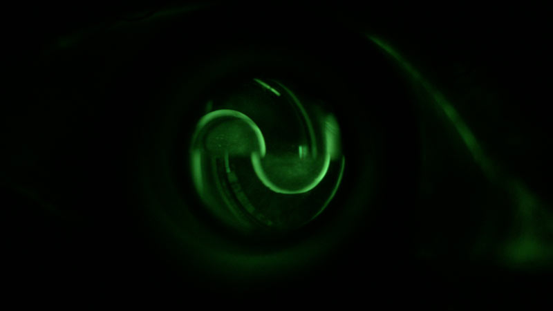
Figure 1: Referral should be considered if intraocular pressure is 24mmHg or more
Diagnosis
Whether in the community as part of a referral refinement scheme, or within the hospital eye service, diagnosis of ocular hypertension or chronic open angle glaucoma requires the following tests to have been carried out:
- Visual field assessment using standard automated perimetry (full threshold).
- Optic nerve assessment and fundus examination using stereoscopic slit lamp biomicroscopy, with pupil dilatation. Image capture, at diagnosis for baseline documentation (for example, a stereoscopic optic nerve head image or OCT) is recommended.
- IOP measurement using Goldmann applanation tonometry (not Perkins).
- Peripheral anterior chamber configuration and depth assessments using gonioscopy – where this is not possible, for example, when people with physical or learning disabilities are unable to participate in the examination) van Herick may be used as an alternative.
- Pachymetry of the central cornea.
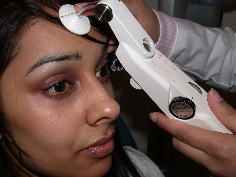
Figure 2: Diagnosis requires slit-lamp mounted and not hand held tonometry
The NICE documents and also a summary of the revision from the College of Optometrists can be viewed here:
http://fplreflib.findlay.co.uk/images/pdf/optician/glaucoma-managing-glaucoma.pdf
http://fplreflib.findlay.co.uk/images/pdf/optician/NICE-guidelines-glaucoma-member-briefing.pdf
http://fplreflib.findlay.co.uk/images/pdf/optician/glaucoma-glaucoma-overview.pdf
http://fplreflib.findlay.co.uk/images/pdf/optician/glaucoma-diagnosis-and-management.pdf
Once you have read through the source material, please attempt the six multiple choice questions;