SeeAbility’s Children in Focus campaign has transformed the delivery of eye care for children with disabilities. Evidence generated over six years of eye examinations in special schools1 underpin both the development of a framework for the provision of eye care in special schools in England2 and the rationale for a national programme of eye care underpinned by the NHS Long-Term Plan.3
This article provides an overview of NHS England’s commitment to commission a national programme of eye care for children with learning disabilities in special schools. It also reviews the epidemiology of a range of conditions that may lead to learning disability, outlining the ocular and systemic associations in each case.
Learning disability and risk to eye health
Mencap defines a learning disability as reduced intellectual ability and difficulty with everyday activities, for example household tasks, socialising or managing money, and which affects someone for their whole life. However, with the right support, people with learning disabilities can and do lead independent lives. Calculated using learning disability prevalence rates from Public Health England (2016) and population data from the Office for National Statistics (2019), the estimated prevalence of learning disabilities in children in the UK is 2.5%. A combination of higher survival rates following premature birth, improved healthcare provision and longer life expectancy are together resulting in a rise in the number of people with learning disabilities.
In England, currently, it is estimated that 79% of children with severe learning disabilities and 81% of children with profound and multiple learning disabilities attend special schools. This amounts to more than 120,000 children.4
Many people with a learning disability may have an associated general health diagnosis which increases the risk of ocular or visual problems. Adults with a learning disability are 10 times more likely to have an eye or vision problem,5 and for children this increases to a staggering 28 times. By contrast, sight problems are comparatively rare in the general paediatric population.6 Due to this increased risk of both visual and ocular problems, it is important that regular and tailored eye care is available and accessible for all children with a learning disability.
Communication difficulties and diagnostic overshadowing (behaviours caused by visual problems being mis-attributed to a learning disability) compound the elevated risk of ocular and visual problems leading to an increased likelihood of significant undiagnosed and untreated visual problems and marked inequality in the provision of eye health care as compared to that for the general population. There is evidence that GOS eye care is only accessed by around 10% of children attending special school1 and that parents may believe eye care is only possible if a child is able to co-operate and can read.7 There is also evidence that, even when eye care is accessed, many of the 31% of children who need refractive correction do not go on to wear spectacles with any success.8 To address this, the model proposed by the SeeAbility framework paper includes spectacle dispensing, fitting and ongoing support with adaptation as part of the service in school.
Preventative eye care measures in this high-risk population are also essential. The introduction of a standardised and systematic programme of eye care within special schools will address existing health inequalities for this high-risk group to access regular eye examinations and spectacles.
Visual screening not appropriate for children with learning disabilities
The UK National Screening Committee (NSC)10 and Public Health England9 recommend orthoptic-led vision screening for all children at school entry (aged four to five years). This is, essentially, to identify amblyopia and any concurrent refractive error and strabismus. The primary aim is to identify and correct reduced vision in such cases. The National Visual Screening Programme adopts the assessment of monocular vision as the main measure of visual function, with a score of 0.2 logMAR acuity in each eye (measured with Keeler Acuity Cards) as the recommended level required to ‘pass’ the screening process.
In special schools, however, the provision of this type of screening is, at best, patchy.8 More importantly, it is doubtful whether such screening methods are suitable for children with complex needs, as many children cannot be assessed using these visual acuity tools.
SeeAbility’s research has found that 85% of children attending their special school service (across all age groups) would either be unable to perform or would fail the vision screening test as set out in NSC guidelines.10 Only 0.6% of the school entry age group passed vision screening. Every child seen by the service was assessed for their presenting level of vision. Limited engagement during an assessment was often followed up with repeated attempts, in classrooms, sensory rooms, and even outdoors according to where a child was most at ease (figure 1). All school entry age, children were seen by an orthoptist and optometrist.
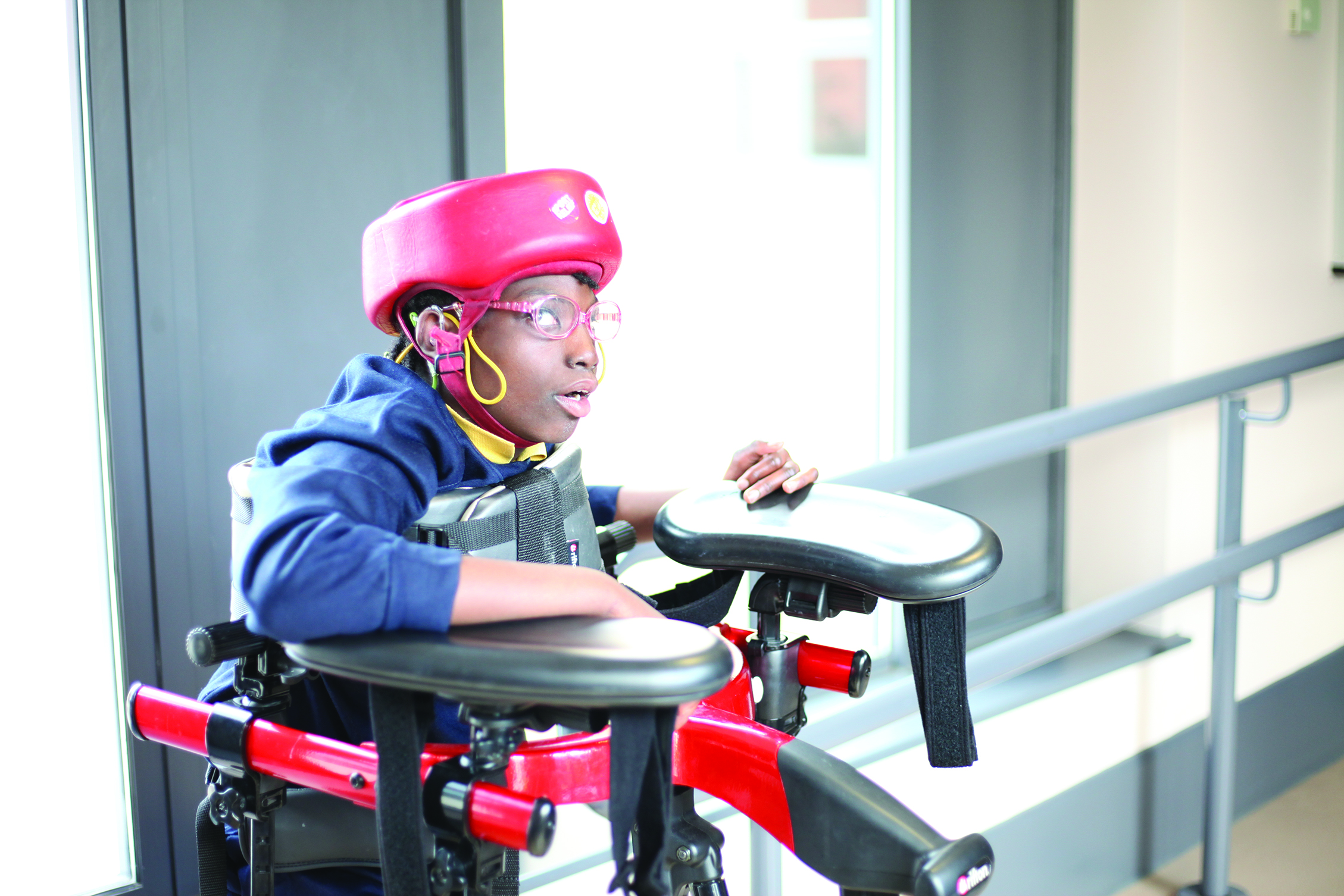 Figure 1: Karen had an eye examination in school supported by her teachers. Her protective helmet was no obstacle to her spectacle wear
Poor uptake and access to GOS Sight Tests
While children in full-time education in England are entitled to regular NHS sight tests, there is strong evidence that children and young people with learning disabilities have problems accessing community eye care services.6,10,11 Any visual difficulties they may be experiencing are therefore often overlooked. This may be due to the difficulty in self-reporting, or diagnostic overshadowing. Even when a carer knows a child very well, a significant visual problem or change in vision can be missed or instead considered to be part of their learning disability.
A high level of unmet eye care within the special schools’ population has been reported in a number of studies,6,8,11 both in community and hospital eye care services. This is echoed by SeeAbility data that reported some 44% of children assessed had received no previous eye care.1 This figure remained remarkably constant for all age groups, even children of secondary school age. Few children (10.7%) had accessed a community eye care practice for a routine sight test. Much more common was for children to be seen in busy hospital eye clinics for routine sight tests instead of community-based alternatives due to a lack of appropriate and specialist service.
Causes of learning disability and their systemic associations
With an estimated 2.5% prevalence of children with learning disability, there are approximately 299,000 children (aged zero to 17 years) with a learning disability in England, of which 101,000 children are in the zero to five years age range.12 Often the ocular anomalies that present in a person with a learning disability may be associated with an underlying general health condition or syndrome.
Prematurity, low birth weight and cerebral palsy
Prematurity is defined as birth before 37 weeks’ gestation and low birth weight as less than 2.5kg. Globally, approximately 10% of babies are born prematurely. Most pre-term births (over 90%) survive without any neurodevelopmental impairment.13 Moderate or severe neurodevelopmental impairment presents in 2.7% of babies born prematurely, while the prevalence of mild neurodevelopmental impairment is a little higher, at an estimated 4.4%.14 Prematurity and low birth weight each, independently, increase the risk of both learning disabilities and ocular problems, with the risk increasing with lower gestational age and birth weight.
Premature babies are more likely to develop cerebral palsy (CP) and hence the systemic, ocular and visual issues associated with it (figure 2). Other common systemic associations of premature birth include pulmonary and hearing problems.  Figure 2: Lana has cerebral palsy. She wears her glasses well and enjoys playing with a range of sensory objects
As a result of brain injury before, during or shortly after birth, it is estimated that one in 400 babies born in the UK have CP.15 It is more likely to occur with a premature or difficult birth. CP may have a number of causes, including:
- Maternal infections during pregnancy, such as cytomegalovirus, rubella, chickenpox and toxoplasmosis)
- Birth hypoxia
- Intracranial haemorrhage or traumatic insult
- Very low blood sugar after birth
- Neonatal infection, such as meningitis
CP can be classified into four categories:
- Spastic; muscle weakness or stiffness, and possibly hemiplegia, diplegia or quadriplegia
- Athetoid; compromised muscle tone resulting in involuntary spasm
- Ataxic; problems with balance and co-ordination
- Mixed; used to describe any combination of the first three
categories
Motor signs vary widely in severity, and may include delay in achieving the expected development milestones, such as the ability to sit upright by eight months or walk by 18 months. Hypotonia (weakness of the arms or legs), fidgety, jerky, clumsy or random movements, muscle spasms, tremors, and walking on tip-toes all feature as characteristic motor skill deficiencies in CP.
Other clinical signs include drooling, gastro-oesophageal reflux disease, constipation, sleep disturbances, epilepsy, hearing loss, scoliosis (curvature of the spine), hip dislocation, difficulties with feeding, and delayed speech and language development. About half of people with CP will have a learning disability, around 20-25% will have epilepsy, and at least 60% exhibit speech and language difficulties.15
Cerebral palsy presents with a high prevalence of visual problems, and 75% of people with CP will have some degree of ocular or visual deficiency. For around 10%, there is a severe visual impairment.16
Down’s syndrome
Trisomy 21, or Down’s syndrome (DS), is caused by a duplication of all or part of chromosome 21, making three copies of the chromosome rather than the usual two copies.
Incidence is approximately one in every 700 births,17 this increasing with maternal age. Physical signs include:
- Short neck
- Excess skin at the back of the neck
- Small head, ears and mouth
- Flat nasal bridge
- Small, often low-set, ears
- High arched palate
- Short fingers
Delays in rolling over, sitting up, crawling, and walking are common in children with DS. This is due to hypotonia, the term for poor muscle tone and low strength. The most common birth defect, seen in 50% of babies with DS, is congenital heart disease (CHD).18 In turn, CHD can lead to high blood pressure particularly affecting pulmonary circulation, an inability of the heart to pump blood effectively and efficiently, and cyanosis (blue-tinted skin caused by reduced oxygen in the blood).
Up to 75% of people with DS have some hearing loss. The incidence increases significantly with age, so that over 90% of people with DS over the age of 50 will have significant hearing loss. People with DS are also much more likely to contract disease associated with immunodeficiency, such as blood disorders including leukaemia, anaemia, polycythaemia, as well as hypothyroidism.19 Regular screening for all these conditions should be part of routine healthcare for people with DS.
Up to 13% of children with DS have epilepsy, this rising to 50% of people aged over 50.20 Seizures are usually well managed with appropriate medication.21 Learning disability in DS is rarely severe. Typically, people with DS have just a mild to moderate disability. Common cognitive and behavioural traits include a short attention span, poor judgment, impulsive behaviour, slow learning and delayed language and speech development. There is also a higher incidence of autism, with around 6 to 7% of children with DS having a dual diagnosis.22
Premature ageing is a characteristic of adults with DS, as is dementia, memory loss, and impaired judgment similar to that experienced with Alzheimer’s disease.23 Other medical concerns associated with ageing DS people include; high cholesterol, obesity, metabolic syndrome, diabetes and early menopause. In contrast, arteriosclerosis, hypertension and solid tumour cancers (for example, breast cancer) have a much lower incidence in people with DS than in the general population.24
Autism spectrum disorders (ASD)
The National Autistic Society defines autism as: ‘a lifelong developmental disability that affects how people perceive the world and interact with others.’25 A clinical diagnosis is made when there is evidence of all of the following:26
- Communication impairment
- Social impairment
- Repetitive stereotyped behaviour
People with autism are often described as being either hypo-responsive or sensory seeking. These different, altered responses to sensory stimuli affect behaviour profoundly.
The prevalence of autism in the UK is around 2% in men and 0.3% in women. It is important to note that autism is not itself a learning disability. However, it can often co-exist with a learning disability. It is estimated that some 50 to 70% of people with an autistic spectrum condition will also have a learning disability.27 Conversely, around 17% of the learning disabled population will also have a diagnosis of autism.22
The prevalence of autism increases with the severity of learning disability, and autism is now the most common primary diagnosis in the special school population.28 Because people with autism are more likely to have a learning disability, they are also more likely to have the general health conditions associated with their learning disability. Other medical disorders associated with autism include; epilepsy, hearing loss, hydrocephalus, foetal alcohol syndrome and CP.29 Depression, anxiety and obsessive compulsive disorder are all more common in people with autism.30
Hydrocephalus
Hydrocephalus describes a build-up of cerebrospinal fluid inside the skull. This can be congenital or acquired. Spina bifida, a neural tube defect where there is incomplete closure of the tissues around the spinal cord, occurs in around 0.1% of babies, most of whom will have hydrocephalus and neurodevelopmental impairment.31
Presentation of congenital hydrocephalus includes an unusually large head, muscle spasms, poor feeding, irritability and drowsiness. Early in childhood, children often have speech difficulties, poor attention, poor co-ordination and epilepsy, leading to a diagnosis of learning disability.
Ocular Associations of Learning Disability
Within the SeeAbility cohort of children, diagnoses of the nature of the disability was known for 90% of children.1 Of these, 51.9% were known to have autism, and 48.1% had another diagnosis, such as cerebral palsy, Down’s syndrome or global developmental delay (GDD). The term global developmental delay is used to describe a child who is slow in reaching two or more milestones in all areas of development.
A number of ocular manifestations were observed during the course of the eye examinations in special schools, and these will now be considered in turn, and any specific disease association discussed.
Nearly half of the children tested had a significant problem with their vision. A pathological disorder was recorded in 7.6% pupils. These included:
- Blepharitis (four cases)
- Conjunctivitis (five cases)
- Keratoconus (four cases)
- Cataract (12 cases)
- Optic disc anomalies (four cases)
- Retinal abnormalities (seven cases)
Vision or acuity loss
Formal vision and acuity checks can be challenging for children with learning disability, even when using objective, preferential-looking techniques (techniques to assess visual function will be discussed in later articles. That said, a quantifiable measure of vision using formal vision tests was possible in about 60.5 % of the cohort of children across all ages. However, for a large proportion of the children (33%) co-operation and comprehension proved too challenging to reliably determine their level of vision.
People with CP have a high incidence of reduced visual acuity. With increasing severity of CP, the likelihood of poor acuity is greater. LogMAR acuities in children with CP have been shown to be, on average, two Snellen-equivalent lines less than those of age-matched controls.16 This may be multifactorial, with possible causes being cerebral visual impairment, imperfect vestibulo-ocular reflex compensation and limited head control.
The high incidence of refractive and strabismic amblyopia may be the most common causes of reduced acuity in children with DS. However, there is some evidence of cortical differences in people with DS. As these changes are similar to those seen in non-DS amblyopes, it is not clear if the differences are a result of the amblyopia or a specific characteristic of DS. Regardless, early and effective refractive correction is strongly advocated in people with DS to avoid, or at least minimise, the risk of reduced acuity.32
Refractive error
Within the overall SeeAbility cohort, 31.5% needed spectacles and 12.9% had spectacles fitted for the first time. Ninety-six point three percent of these children had their glasses fitted in school, highlighting the importance of spectacle dispensing to a sight testing service within special schools.10
Myopia of greater than -0.50DS in either eye was seen in 22.3% of students, nearly double the 11.7% prevalence in the general paediatric population.33 Significant uncorrected high myopia (>-6.00DS) and pathological myopia (> -13.00DS) were also detected in both primary and secondary school age groups.
Similarly, mild hypermetropia (≥+2.00DS) in either eye was seen in 15.2% of pupils, more than 3 times the 4.7% prevalence in the overall childhood population. Astigmatism (defined as ≥ 1.00DC in either eye and present in isolation or co-existing with myopia or hyperopia) was measured in 28.6% of children, nearly double the 14.9% prevalence within the overall childhood population.34
Pre-term children, both with and without retinopathy of prematurity (ROP), are more likely to develop myopia and astigmatism.35,36
A failure of the emmetropisation process is believed to be the cause of the high prevalence (around 60 to 80%) of refractive error in children with CP. The more severe a person’s gross motor impairment, the higher their level of refractive error is likely to be. Astigmatism is higher in magnitude and its prevalence rises with increasing degree of intellectual impairment.37
The prevalence of refractive errors is high in children with Down’s syndrome. At birth, there is no significant difference in the prevalence of refractive error between those with DS and controls.38 However, a longitudinal study by Woodhouse et al showed a higher incidence of refractive error in children with DS when compared to a control group.39 Children with DS do not undergo the emmetropisation found in the general population and refractive error often increases with age.40 Also, oblique astigmatism is more common, with both the magnitude and axis likely to change over time. 41
Refractive errors have been found to be more common in people with autism with 20 to 30% reported to have significant error, and with increasing levels of astigmatism consistently reported.42
Strabismus
Two percent of the general paediatric population are reported to have strabismus.43 Analysis of the SeeAbility data shows that binocular vision anomalies were found in 25.2% of pupils, with strabismus being the most reported, followed by nystagmus and a lack of smooth pursuit eye movements.10
Strabismus is much more common in babies born before 28 weeks gestation. 14% of babies born before 28 weeks have strabismus, as compared to around 2% of the overall childhood population.43
Strabismus is also very common in CP, occurring in over 50% of people with the condition.44 Either exotropia or esotropia (rarely accommodative) are often present, and there is a high prevalence of both nystagmus and incomitant strabismus, often linked to trauma to the head.45 Referral for surgery should always be discussed as, despite the poor prognosis in terms of any likely restoration of binocular vision, the psychosocial benefit of an improved appearance can significantly add to quality of life.46
A particularly high prevalence of esotropia (35%) has been reported in people with DS. While esotropia may be associated with hyperopia and have a variable accommodative element linked to a poor accommodative ability, it is not uncommon to find a myope with an eso-deviation. This is a rare finding in the general population. The onset of strabismus in DS is at a later age than typical in the general population, averaging at 4.5 years. This highlights the need to monitor and assess binocular status at school entry age.47 Prominent epicanthal folds, common in those with DS and causing a pseudostrabismus, may affect the practitioner’s ability to assess the presence of strabismus.
Strabismus has been reported to be highly prevalent in children with autistic spectrum disorder (ASD), at least twice as high as that within the general population.48
Eye movement abnormalities
Coordinated, controlled eye movements are essential to perceive and react to the visual environment. Eye movements provide essential visual sensory information to support cognitive development and learning. Conversely, deficits in the control of both voluntary and involuntary eye movements impact upon a child’s ability to understand different types of motion, objects and conditions. Eye movement deficits are more frequently found in children with learning disabilities.49
Twelve to 25% of children with Down’s syndrome will have nystagmus.32,50
Eye movement abnormalities are common with CP, and both smooth pursuits and saccades may be affected. Four to 10% of children with CP will have nystagmus.45,51-53
Eye movements have been shown to be atypical in people with autism,54 although it is difficult to gauge the impact upon visual processing. While gross control of fixation is intact, deficits in smooth pursuit movements in children and young adults with ASD mean that maintaining controlled and accurate fixation is difficult.
Accommodative problems
Due to the high incidence of accommodative problems in the learning-disabled population, it is important to always assess accommodation, and a near addition in the form of reading glasses or bifocal or varifocal correction may be needed at any
age, including in childhood (figure 3). 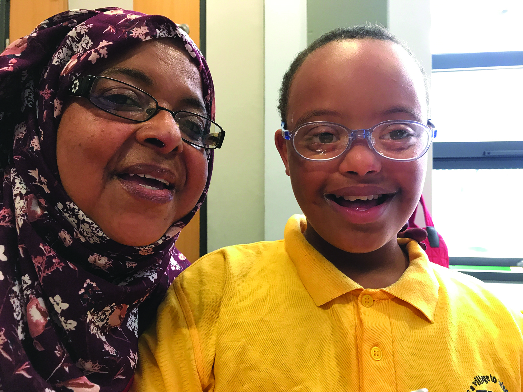 Figure 3: Nasir with his mother, soon after being fitted with bifocals
Dynamic retinoscopy techniques, as well as subjective measurement of accommodative ability, are recommended. It is also worth bearing in mind that lower levels of hyperopia than typically require correction may need to be corrected for children
and younger adults with poor accommodation.
Fifty to 60% of children with CP have been shown to have significantly reduced accommodation,53-55 and a reading addition may need to be considered irrespective of age. Restricted control of head movements can make the use of multifocal spectacles impractical and may necessitate the prescribing of designated single vision prescriptions for different tasks. Reduced accommodative responses are strongly associated with more severe motor and intellectual impairment. Accurate and task-specific prescribing and dispensing of refractive correction for people with CP is critical to optimal visual performance. For example, it is essential to consider the working distance for any screen-based communication technology, any head rests and supports, and the extent of control of head movement.
There is good evidence for the presence of low amplitude and poor control of accommodation in the majority, around 80%, of children with DS.29,36,55,56
Accommodative problems are three times more prevalent in people with autism than in the general population.55
Keratoconus
Relatively rare in the general population with a prevalence of 0.27%,61 keratoconus is much more commonly found in people with learning disabilities.
Some 5.5% of people with Down’s syndrome will develop keratoconus.62 Early detection and referral are essential to halt progression (using corneal collagen cross-linking, or CXL) at as early a stage as possible before the cornea becomes too thin.63 CXL is usually carried out under local anaesthesia, although general anaesthesia may be preferred if anxiety is high for a young person with a learning disability.
Cataracts
Detection, monitoring and timely referral for cataract surgery is essential for children with a learning disability.
Diabetic retinopathy screening
It is of utmost importance that children with diabetes and learning disabilities are seen for regular retinal assessment, with dilation when possible, either as part of the Diabetic Retinal Screening Programme or within a community or hospital setting where they may be more comfortable and cooperative.
Retinopathy of prematurity (ROP)
The prevalence of ROP, a proliferative retinal vascular disease affecting premature infants, is about 5% in babies born in the UK before 32 weeks.14 The clinical spectrum of ROP ranges from spontaneous regression to bilateral retinal detachment and complete functional blindness. Between these two extremes lies the form of ROP which is amenable to treatment with laser photocoagulation, anti-vascular endothelial growth factor drugs or surgery. The risk of developing ROP and its severity increases with reducing gestational age, decreasing birth weight, the presence of intraventricular haemorrhage and with pulmonary disease.14,64
People with a learning disability who have a history of ROP are at higher risk of developing retinal detachment and glaucoma.
Optic nerve disorders
Fifteen to 40% of people with congenital hydrocephalus will have optic atrophy. The prevalence is likely lower in the younger population thanks to better early management of the condition in recent years. There is an increased incidence of strabismus, eye movement anomalies and refractive error associated with optic atrophy.59
Disc hypoplasia, pallor and high cup-to-disc ratios are more common in people with CP, the prevalence of pallor as high as 35%.40
Visual field defects
Within the SeeAbility cohort, 2% of children were found to have a significant visual field defect. The majority of these were hemianopias and 22% were previously undiagnosed.
Visual field defects are common in people with CP. It is important to detect such a defect and clearly communicate to families, carers and teachers its nature in order to support rehabilitative intervention and to maximise mobility. The positioning of teachers and carers, headrests on wheelchairs, and of the child within a classroom all depend upon a good understanding of any visual field deficit.
Hemianopias (and other visual field defects) are associated with CP, most frequently with hemiplegic CP, with one study reporting an incidence of 64%.45
Visual processing disorders and cerebral visual impairment
Cerebral visual impairment (CVI) is the consequence of neurological injury, resulting in a huge spectrum of possible clinical presentations of visual impairment. Visual function may be minimally affected with only slightly reduced visual acuity and no other apparent processing issues, or cerebral damage may be severe enough to result in no useful visual function at all. Widely variable visual abilities are observed and the extent is heavily influenced by other sensory activity. Visual agnosia, for example, is much worse with fatigue or engagement with other sensory activities. Problems with movement detection and with eye movement control may all cause variable visual deficiency. Repeated and regular assessment is important, as neuroplasticity can result in changes to the clinical presentation of CVI over time.65
As the visual cortex is a part of the cerebral cortex, and so much of both cognitive development and mobility is tied to the body’s ability to interpret and process visual information, a high proportion of children with CP (documented as 60 to 70%) also have some degree of CVI.51,66
Individuals with autism report that they perceive the world differently. There generally seems to be a heightened sensitivity to local, detailed percepts and a weaker recognition of the global ‘big picture’; the meaning or context of a stimulus. This makes visual crowding and complex tasks more challenging.67,54 In the absence of severe visual impairment, there is no evidence that we should expect reduced visual acuity in people with autism.55
Development of a Framework for Special Schools Eye Care in England
With an increasing evidence base to address the inequalities in access and uptake of eye examinations for children with learning disability, a standardised and comprehensive approach to improving care is required.
SeeAbility’s framework for the provision of eye care in special schools in England68 was developed in association with the Association of British Dispensing Opticians, the British and Irish Orthoptic Society, the College of Optometrists, LOCSU and the Royal College of Ophthalmologists, and endorsed by the Clinical Council for Eye Health.
The framework recommends clinical protocols for regular, full routine eye examinations to be offered to all students in their special school environment. There are recommendations for pre-assessment questionnaires, a list of required equipment, and templates for reports to parents, carers and teachers. The framework advocates the provision of routine eye care; including refraction, eye health checks and spectacle dispensing, and importantly the effective dissemination of visual status findings to parents and all health and education specialists supporting children with learning disabilities.
The Path to Change
With a staggeringly low uptake of free NHS eye examinations among children with learning disability, together with a much higher prevalence of visual related problems, it has become clear that the NHS General Ophthalmic Service is not fit for children with complex needs in England. With a strong evidence base of the prevalence of ocular problems, and a framework of clinical protocols, there is an urgent case for the delivery of a consistent and comprehensive nationwide programme of eye care. NHS England is now fully committed to delivering this in order to meet the targets of the Long-Term Plan, and to maintain its clinical focus on addressing the health inequalities experienced by people with learning disabilities and autism.
Along with NHS England’s commitment to the inclusion of sight testing in special schools, Public Health England has recently revised its service specification and now recommends the framework model as an appropriate alternative to school entry screening for this population.9 Children with the most profound learning disabilities, and hence the most likely to have associated visual problems, are more likely to attend special schools. Special schools, where children are familiar and comfortable with their surroundings and where their educational development, function and attainment are fully supported, are ideal places to conduct eye examinations (figure 4). Also, parents have expressed a strong preference for an in-school model of eye care and spectacle dispensing.69 Follow-up examinations, necessary where there have been challenges initially, spectacle fitting and checks, and sharing of information with the multidisciplinary team in school are all amenable to the school environment. 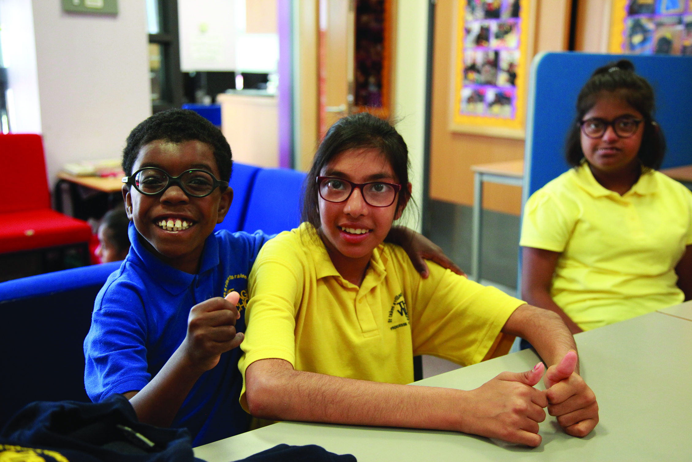 Figure 4: Spectacles can be conveniently dispensed in the classroom to meet the high prevalence of refractive error among children with learning disabilities
A Call to Action
All practitioners interested in becoming involved with the provision of eye examinations and spectacle dispensing in special schools can learn more on the newly set up specialist Learning Disabilities Eye Care Interest Group. For further information, go to www.linkedin.com/posts/seeability-eye-care_specialschoolseyecare-activity-6646479658239963137-719P.
While children attending special schools will be able to access regular eye care with the implementation of the new NHS special schools eye care service, encouraging the uptake of routine primary eye care and raising awareness of the risks to visual function for both adults with learning disabilities and children attending mainstream schools and their carers, is an important responsibility for all eye care practitioners.
Most importantly, correcting refractive error, even for children with poor vision or visual processing difficulties, and the importance of spectacle dispensing (figure 5) with regular fitting checks and repairs cannot be emphasised enough. There is good evidence that this is beneficial to optimise visual performance even in the presence of significant CVI.70 Figure 5: Kiyana enjoying trying on a range of frames!
There are a wide range of resources available free to download from www.seeability.org (including easy to read fact sheets on many eye conditions) to help your practice to support people with learning disabilities with their eye care. The ‘Telling the Optometrist About Me’ form can be used as a pre-assessment questionnaire, and the ‘Results of my Eye Test’ can be used to feed into a person’s care strategy regarding their visual functions and needs.
Children with learning disabilities should have equal access to eye care across primary and secondary care, from refractive correction through to invasive surgical procedures, to enable them to play an active part in society and to access the education, employment and leisure available to others.
Sonal Rughani is a specialist public health optometrist and works with SeeAbility as well as acting as a specialist optometrist for the RNIB in London.
Lisa Donaldson is clinical lead for SeeAbility’s Special Schools Service, and primary care clinical lead for NHS England’s Special Schools Eye Care Programme, Visiting Lecturer at City, University of London and University of
Hertfordshire.
References
- Donaldson LA, Karas M, O’Brien D, Woodhouse JM. Findings from an opt-in eye examination service in English special schools. Is vision screening effective for this population? Awadein A, editor. PLoS One. 2019 Mar;14(3):e0212733.
- SeeAbility, RCOphth, BIOS, LOCSU, CollegeOptoms A. Framework for Special Schools Eye Care. 2016.
- Cheater S. The NHS Long-Term Plan. International Journal of Health Promotion and Education. 2019.
- Gov.uk. 2019 UK Gov data on SEN. https://assets.publishing.service.gov.uk/government/uploads/system/uploads/attachment_data/file/804374/Special_educational_needs_May_19.pdf. 2019
- Emerson E, Robertson J. The Estimated Prevalence of Visual Impairment among People with Learning Disabilities in the UK. RNIB SeeAbility [Internet]. 2011;35. Available from: http://www.rnib.org.uk/aboutus/Research/reports/2011/Learn_dis_small_res.pdf
- Das M, Spowart K, Crossley S, Dutton GN. Evidence that children with special needs all require visual assessment. Arch Dis Child. 2010;95(11):888–92.
- Donaldson L, Subramanian A, Conway ML. Eye care in young children: a parent survey exploring access and barriers. Clin Exp Optom. 2018;
- Woodhouse JM, Davies N, McAvinchey A, Ryan B. Ocular and visual status among children in special schools in Wales: The burden of unrecognised visual impairment. Arch Dis Child. 2014;99(6):500–4.
- Public Health England. Child Vision Screening Service Specification. https://www.gov.uk/government/publications/child-vision-screening/service-specification. 2019.
- Hall DMB, Elliman D. Health for all children (rev. 4th ed.). [Internet]. Health for all children (rev. 4th ed.). 2006. Available from: http://ovidsp.ovid.com/ovidweb.cgi?T=JS&PAGE=reference&D=psyc5&NEWS=N&AN=2007-00943-000
- Pilling RF, Outhwaite L. Are all children with visual impairment known to the eye clinic? Br J Ophthalmol [Internet]. 2017 Apr;101(4):472–4. Available from: http://bjo.bmj.com/lookup/doi/10.1136/bjophthalmol-2016-308534
- E. E. Household deprivation, neighbourhood deprivation, ethnicity and the prevalence of intellectual and developmental disabilities. J Epidemiol Community Heal. 2012;66:218–24.
- Blencowe H, Lee ACC, Cousens S, Bahalim A, Narwal R, Zhong N, et al. Preterm birth-associated neurodevelopmental impairment estimates at regional and global levels for 2010. Pediatr Res. 2013;
- O’Connor a R, Wilson CM, Fielder a R. Ophthalmological problems associated with preterm birth. Eye (Lond). 2007;21(10):1254–60.
- Odding E, Roebroeck ME, Stam HJ. The epidemiology of cerebral palsy: Incidence, impairments and risk factors. Disabil Rehabil. 2006;
- Ghasia F, Brunstrom J, Gordon M, Tychsen L. Frequency and severity of visual sensory and motor deficits in children with cerebral palsy: Gross motor function classification scale. Investig Ophthalmol Vis Sci. 2008;49(2):572–80.
- Hobson-Rohrer WL, Samson-Fang L. Down’s syndrome. Pediatr Rev [Internet]. 2013;34(12):573–4. Available from: http://pedsinreview.aappublications.org/cgi/doi/10.1542/pir.34-12-573
- Stoll C, Dott B, Alembik Y, Roth MP. Associated congenital anomalies among cases with Down’s syndrome. Eur J Med Genet. 2015;
- Henderson A, Lynch SA, Wilkinson S, Hunter M. Adults with Down’s sydrome: The prevalence of complications and health care in the community. Br J Gen Pract. 2007;
- Ulate-Campos A, Nascimento A, Ortez C. Down’s syndrome and epilepsy. Int Med Rev Down Syndr. 2014;
- Barca D, Tarta-Arsene O, Dica A, Iliescu C, Budisteanu M, Motoescu C, et al. Intellectual disability and epilepsy in Down’s syndrome. Maedica (Buchar). 2014;
- Emerson E, Baines S. The estimated prevalence of autism among adults with learning disabilities in England. Improv Heal Lives Learn …. 2010;
- Deb S, McHugh R. Dementia among persons with Down’s syndrome. International Review of Research in Mental Retardation. 2010.
- Malt E a, Dahl RC, Haugsand TM, Ulvestad IH, Emilsen NM, Hansen B, et al. Health and disease in adults with Down’s syndrome. Tidsskr den Nor Laegeforening. 2013;
- National Autistic Society. Autism Facts and History. NAS. 2019.
- Singer E. Redefining autism. Nature. 2012;
- Brugha T, Cooper S, McManus S, Purdon S, Smith J, Scott F, et al. Estimating the prevalence of Autism Spectrum Conditions in adults: extending the 2007 adult psychiatric morbidity survey. NHS Inf Cent Heal Soc Care. 2012;
- Gov.uk. 2019 UK Gov data on SEN. https://assets.publishing.service.gov.uk/government/uploads/system/uploads/attachment_data/file/804374/Special_educational_needs_May_19.pdf. 1395.
- Croen LA, Zerbo O, Qian Y, Massolo ML, Rich S, Sidney S, et al. The health status of adults on the autism spectrum. Autism. 2015;
- The National Institute of Mental Health. NIMH » Anxiety Disorders. U. S. Department of Health and Human Services. 2016.
- Spina Bifida Fact Sheet. Natl Inst Neurol Disord Stroke. 2013;
- Tomita K, Tsurui H, Otsuka S, Kato K, Kimura A, Shiraishi Y, et al. [Ocular findings in 304 children with Down’s syndrome]. Nihon Ganka Gakkai Zasshi [Internet]. 2013;117(9):749–60. Available from: http://www.ncbi.nlm.nih.gov/pubmed/24261190
- Morgan IG, French AN, Ashby RS, Guo X, Ding X, He M, et al. The epidemics of myopia: Aetiology and prevention. Progress in Retinal and Eye Research. 2018.
- Hashemi H, Fotouhi A, Yekta A, Pakzad R, Ostadimoghaddam H, Khabazkhoob M. Global and regional estimates of prevalence of refractive errors: Systematic review and meta-analysis. Journal of Current Ophthalmology. 2018.
- Gursoy H, Basmak H, Bilgin B, Erol N, Colak E. The effects of mild-to-severe retinopathy of prematurity on the development of refractive errors and strabismus. Strabismus [Internet]. 2014 Jun 1;22(2):68–73. Available from: https://doi.org/10.3109/09273972.2014.904899
- Zhu X, Zhao R, Wang Y, Ouyang L, Yang J, Li Y, et al. Refractive state and optical compositions of preterm children with and without retinopathy of prematurity in the first 6 years of life. Med (United States). 2017;96(45).
- Saunders KJ, Little JA, Mcclelland JF, Jonathan Jackson A. Profile of refractive errors in cerebral palsy: Impact of severity of motor impairment (GMFCS) and CP subtype on refractive outcome. Investig Ophthalmol Vis Sci. 2010;
- Margaret Woodhouse J, Pakeman VH, Cregg M, Saunders KJ, Parker M, Fraser WI, et al. Refractive errors in young children with Down’s syndrome. Optom Vis Sci. 1997;
- Cregg M, Woodhouse JM, Stewart RE, Pakeman VH, Bromham NR, Gunter HL, et al. Development of refractive error and strabismus in children with Down’s syndrome. Investig Ophthalmol Vis Sci. 2003;44(3):1023–30.
- Watt T, Robertson K, Jacobs RJ. Refractive error, binocular vision and accommodation of children with Down’s syndrome. Clin Exp Optom [Internet]. 2015;98(1):3–11. Available from: http:https://doi.org/10.1111/cxo.12232
- Little JA, Woodhouse JM, Saunders KJ. Corneal power and astigmatism in Down’s syndrome. Optom Vis Sci. 2009;
- Little JA. Vision in children with autism spectrum disorder: a critical review. Clin Exp Optom. 2018;
- Pathai S, Cumberland PM, Rahi JS. Prevalence of and early-life influences on childhood strabismus: Findings from the millennium cohort study. Arch Pediatr Adolesc Med. 2010;164(3):250–7.
- Erkkilä H, Lindberg L, Kallio AK. Strabismus in children with cerebral palsy. Acta Ophthalmol Scand. 1996;
- Fazzi E, Signorini SG, La Piana R, Bertone C, Misefari W, Galli J, et al. Neuro-ophthalmological disorders in cerebral palsy: Ophthalmological, oculomotor, and visual aspects. Dev Med Child Neurol. 2012;54(8):730–6.
- Archer S, Musch D, Wren P, E Guire K, Del Monte M. Social and Emotional Impact of Strabismus Surgery on Quality of Life in Children. Vol. 9, Journal of AAPOS : the official publication of the American Association for Pediatric Ophthalmology and Strabismus / American Association for Pediatric Ophthalmology and Strabismus. 2005. 148–151 p.
- Haugen OH, Høvding G. Strabismus and binocular function in children with Down’s syndrome. A population-based, longitudinal study. Acta Ophthalmol Scand. 2001;
- Kaplan M, Rimland B, Edelson SM. Strabismus in autism spectrum disorder. Focus Autism Other Dev Disabl. 1999;
- Fukushima J, Tanaka S, Williams JD, Fukushima K. Voluntary control of saccadic and smooth-pursuit eye movements in children with learning disorders. Brain Dev. 2005;
- Ljubic A, Trajkovski V, Stankovic B. Strabismus, refractive errors and nystagmus in children and young adults with Down’s syndrome. Ophthalmic Genet. 2011;
- Elmenshawy AA, Ismael A, Elbehairy H, Kalifa NM, Fathy MA, Ahmed AM. Visual Impairment in Children With Cerebral Palsy. Int J Acad Res [Internet]. 2010;2(5):67–71. Available from: http://login.ezproxy.library.ualberta.ca/login?url=http://search.ebscohost.com/login.aspx?direct=true&db=a9h&AN=54352948&site=ehost-live&scope=site
- Li JCH, Wong K, Park ASY, Fricke TR, Jackson AJ. The challenges of providing eye care for adults with intellectual disabilities. Clin Exp Optom [Internet]. 2015;98(5):420–9. Available from: http://dx.doi.org/10.1111/cxo.12304
- Sasmal NK, Maiti P, Mandal R, Das D, Sarkar S, Sarkar P, et al. Ocular manifestations in children with cerebral palsy. J Indian Med Assoc. 2011;
- Little JA. Vision in children with autism spectrum disorder: a critical review. Clin Exp Optom [Internet]. 2018; Available from: https://www.ncbi.nlm.nih.gov/pubmed/29323426%0Ahttp://onlinelibrary.wiley.com/store/10.1111/cxo.12651/asset/cxo12651.pdf?v=1&t=jcm5a7se&s=6615c4262383e358a03eb8eab6e280d3bca3ba32
- Anketell PM, Saunders KJ, Gallagher SM, Bailey C, Little JA. Accommodative Function in Individuals with Autism Spectrum Disorder. Optom Vis Sci. 2018;95(3):193–201.
- McClelland JF, Parkes J, Hill N, Jackson AJ, Saunders KJ. Accommodative dysfunction in children with cerebral palsy: a population-based study. Invest Ophthalmol Vis Sci. 2006;47(5):1824–30.
- Leat SJ. Reduced accommodation in children with cerebral palsy. Ophthalmic Physiol Opt. 1996;16(June 1995):385–90.
- Woodhouse JM, Meades JS, Leat SJ, Saunders KJ. Reduced accommodation in children with Down’s syndrome. Investig Ophthalmol Vis Sci. 1993;34(7):2382–7.
- Aring E, Andersson S, Hård AL, Hellström A, Persson EK, Uvebrant P, et al. Strabismus, binocular functions and ocular motility in children with hydrocephalus. Strabismus. 2007;
- Stewart RE, Woodhouse JM, Cregg M, Pakeman VH. Association between accommodative accuracy, hypermetropia, and strabismus in children with Down’s syndrome. Optom Vis Sci. 2007;
- Godefrooij DA, de Wit GA, Uiterwaal CS, Imhof SM, Wisse RPL. Age-specific Incidence and Prevalence of Keratoconus: A Nationwide Registration Study. Am J Ophthalmol. 2017;
- Shapiro MB, France TD. The Ocular Features of Down’s Syndrome. Am J Ophthalmol [Internet]. 1985 Jun 1 [cited 2018 May 18];99(6):659–63. Available from: https://www.sciencedirect.com/science/article/pii/S0002939414760313
- Mazzotta C, Caporossi T, Denaro R, Bovone C, Sparano C, Paradiso A, et al. Morphological and functional correlations in riboflavin UV A corneal collagen cross-linking for keratoconus. Acta Ophthalmol. 2012;90(3):259–65.
- Prasad G V, K S, M B B S SK. A study of risk of retinopathy of prematurity in low birth weight and premature infants with extended protocols of screening. Int J Adv Res [Internet]. 2018;6(2):2320–5407. Available from: http://dx.doi.org/10.21474/IJAR01/6467
- Leat SJ. Reduced accommodation in children with cerebral palsy. Ophthalmic Physiol Opt [Internet]. 1996;16(June 1995):385–90. Available from: http://www.ncbi.nlm.nih.gov/entrez/query.fcgi?cmd=Retrieve&db=PubMed&dopt=Citation&list_uids=8944183
- Philip SS, Dutton GN. Identifying and characterising cerebral visual impairment in children: A review. Vol. 97, Clinical and Experimental Optometry. 2014. p. 196–208.
- Behrmann M, Thomas C, Humphreys K. Seeing it differently: visual processing in autism. Vol. 10, Trends in Cognitive Sciences. 2006. p. 258–64.
- SeeAbility, RCOphth, BIOS, LOCSU, CollegeOptoms A. Framework for Special Schools Eye Care [Internet]. 2016. Available from: https://www.rcophth.ac.uk/wp-content/uploads/2016/07/Framework-for-Proposed-Special-Schools-Service-Final-ABDO-BIOS-College-of-Optometrists-LOCSU-RcOphth-and-SeeAbility-2.pdf
- Donaldson L, O’Brien D, Karas M. (2) (PDF) Parental views of an in-school eye care service for children with severe learning disabilities. In: British Association of Childhood Disabil. 2018.
- Williams C, Northstone K, Borwick C, Gainsborough M, Roe J, Howard S, et al. How to help children with neurodevelopmental and visual problems: A scoping review. Vol. 98, British Journal of Ophthalmology. 2014. p. 6–12.