So far in the recent CPD series on age-related macular degeneration, the focus has been on assessment and management. In part one (Optician 25.11.22), there was a detailed discussion of the latest theories of the causes of the condition and its many and various risk factors. By knowing these, it is possible to undertake a more focused and detailed history and symptoms to investigate any patient either known to have or suspected to have AMD. Part two of the series (Optician 20.01.23) took a closer look at the investigation of the disease using a variety of techniques, both established and using newer technologies.
This week, we launch a new VRICS CPD exercise aimed at consolidating what we have learned so far. There are 12 cases shown here and, you will find questions relating to each of them.
Retinal Photography
Case 1
This patient was a 67-year-old male attending for a routine eye examination. He was a pseudophake, having had his cataracts removed some five years previously. His last eye examination was just after his surgery and, since then, he had noticed a slow decline in his ability to read smaller print. He is now finding this becoming inconvenient and so has attended today hoping for stronger spectacles.
His current spectacles are as follows:
- R: +0.50/ -0.50 x 180 (6/9)
- L: +0.25 / -0.75 x 175 (6/9)
- Add: +3.00 (N6- R&L) @ 35cm
There is no improvement with pinhole.
What is shown here and how might you best manage the patient?
Case 2
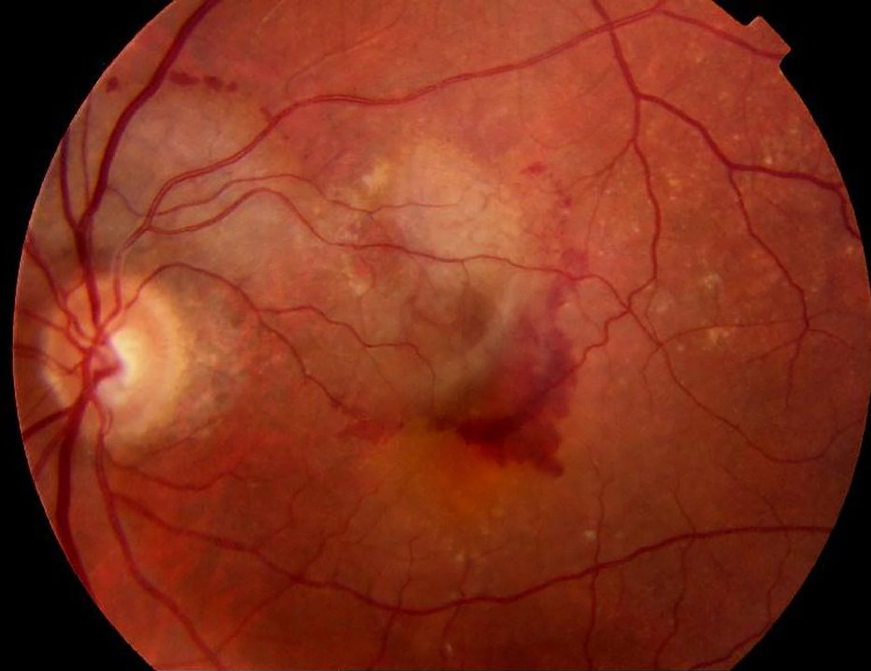
This patient last had an eye examination nine months ago. He is a 70-year-old smoker and had been told at the last eye examination of some ‘age changes at the back of his eyes’. He had been given advice about smoking cessation and diet, and had been issued with an Amsler chart which, he confessed, he had not used. He did, however, feel that there had been some change to his vision in the left eye ‘a week or so’ ago, but had not acted on this. Today, however, he woke to notice a dramatic drop in the vision in this same eye which you measured to be 6/60.
What has happened and what might you do?
Case 3
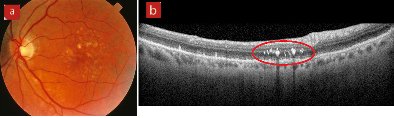
This 71-year-old female attended for her annual eye examination. She had been told a year ago about early AMD changes and given appropriate advice. Her best corrected acuity was 6/9 in each eye. The fundus appearance (figure 3a) appeared very similar to last year. Since last year, your practice has acquired an OCT and a volume scan of the posterior pole was undertaken, one line scan of which is shown in figure 3b.
What does the retinal image show and what are the lesions shown by the OCT scan?
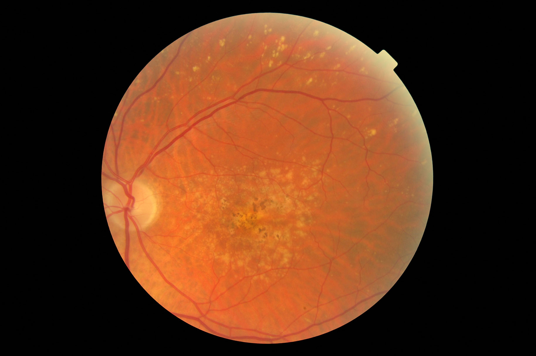
Case 4
This 78-year-old male attended for a low vision assessment, having been certified as eligible for sight impairment registration. His best corrected acuity is 0.9 logMAR right and left, and he can achieve N24 @ 25cm with a +4.00DS add.
What does the retinal image show and how might this man be helped to read his newspaper again?
Optical Coherence Tomography (OCT)
Case 5
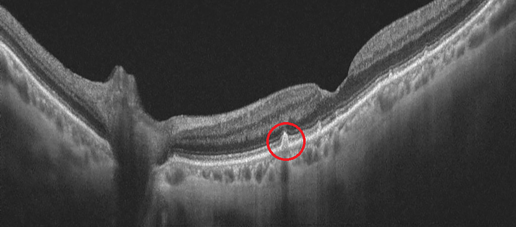
A 75-year-old woman attends for a routine eye test, having previously been told she had dry AMD. Her OCT scan is shown here.
What can be seen in this scan and, in particular, what is shown in the area outlined above in red?
Case 6
This 82-year-old is registered as severely sight impaired, each eye having a best acuity of 1.3 logMAR.
What can be identified around the macula area in the scan?
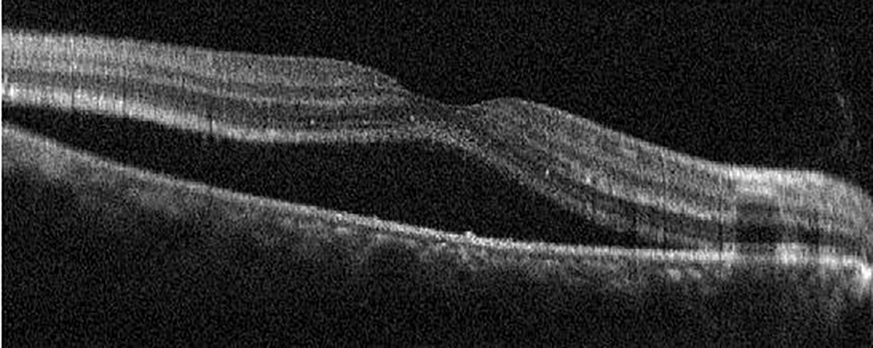
Case 7
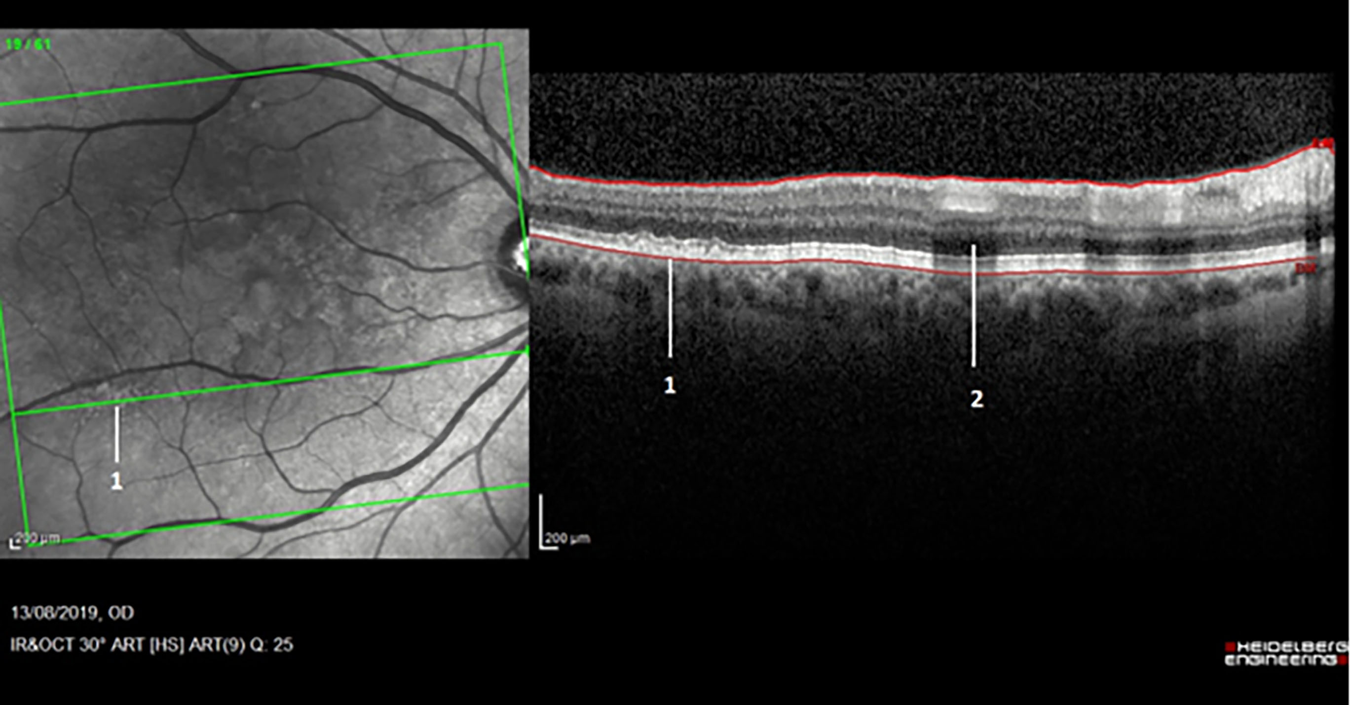
A 61-year-old female smoker attends for a routine eye examination. Her corrected acuities are 6/5 R&L (N4.5 with a +2.25DS add). You undertake a scan of her posterior pole and decide to discuss your findings with her. She asks about two things (marked 1 and 2) on the scan image.
What might these two appearances signify?
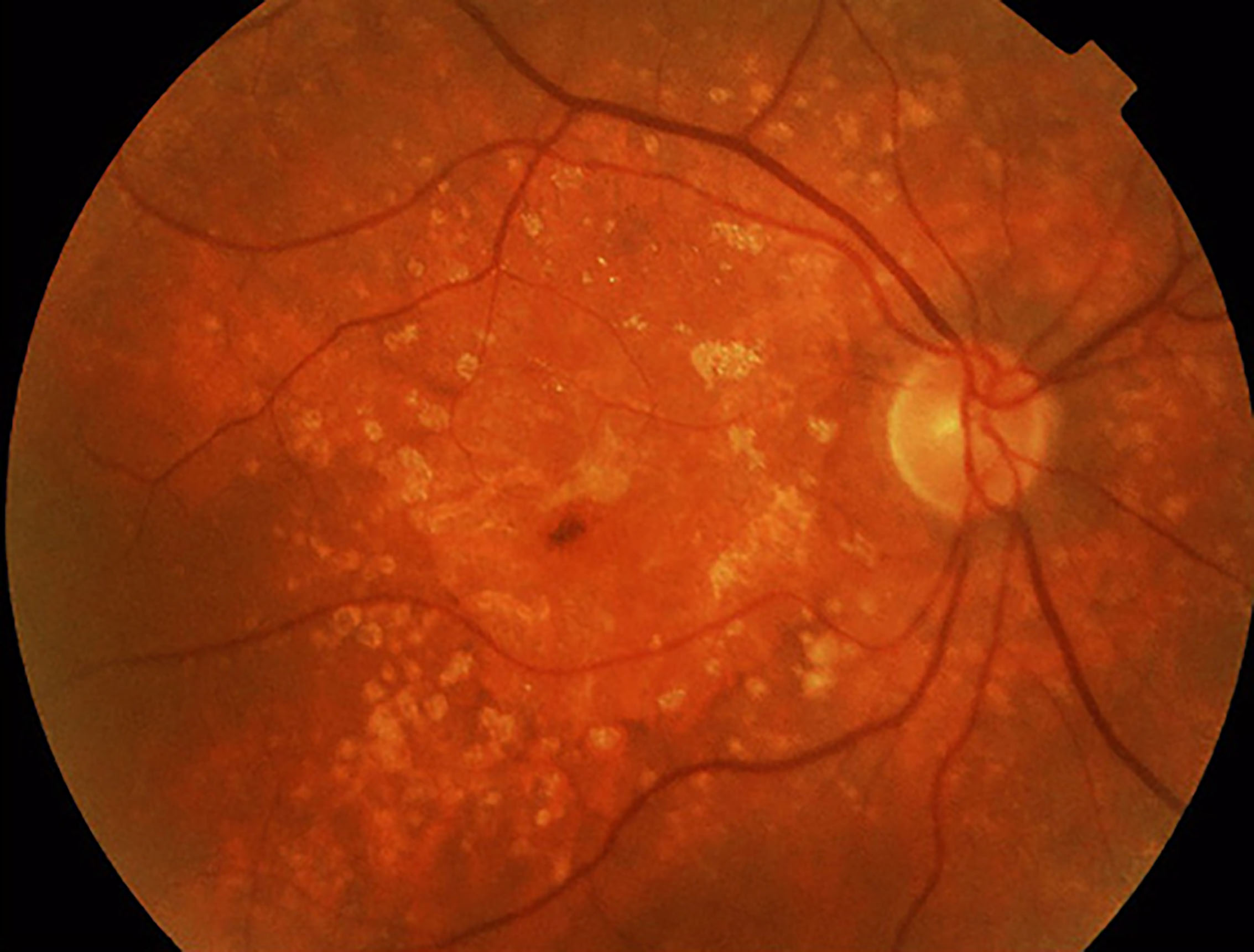
Case 8
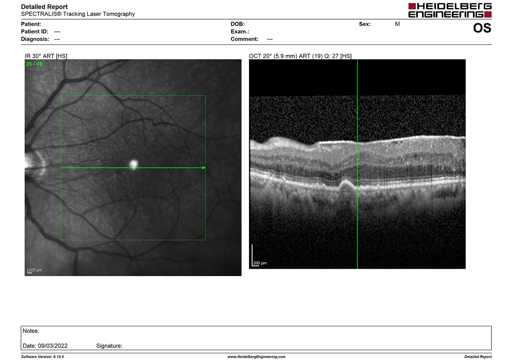
A 78-year-old male myope, with a best corrected acuity of 0.3 logMAR in the left eye has noticed a gradual reduction in the quality of his vision over the past year. His notes show that, around five years ago, he had been referred for a visual disturbance and a posterior vitreous detachment had been confirmed at that time.
What is the bright spot on the left en face image and also what can be seen on the line scan on the right?
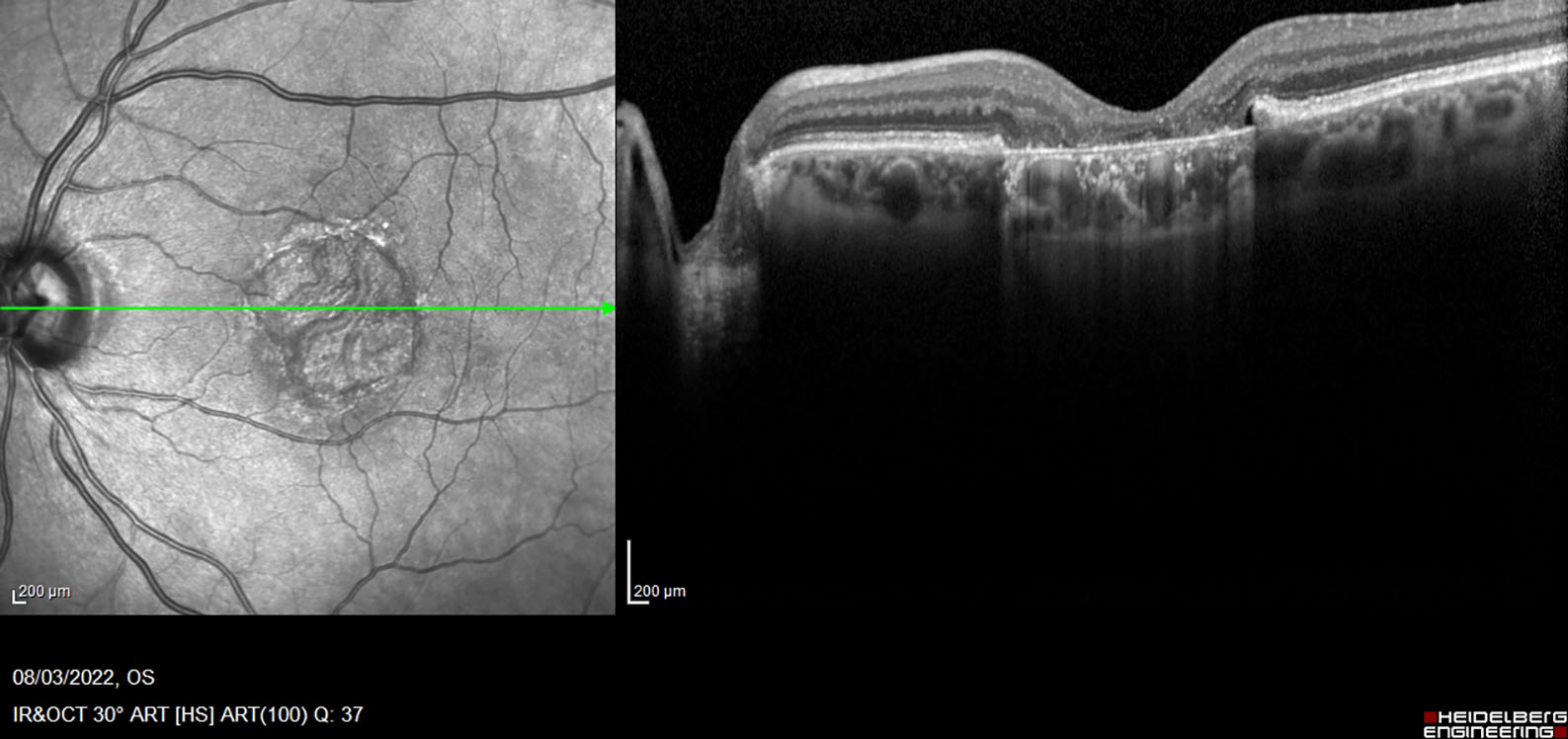
Case 9
A 68-year-old female, to whom you had issued an Amsler chart some eight months ago, attends for an appointment within 24 hours of telephoning your practice to say she had noticed distortion of the grid that morning. This had not been noticed previously.
What is shown in this scan and what might be your management of the patient?
Advanced Technologies
Case 10
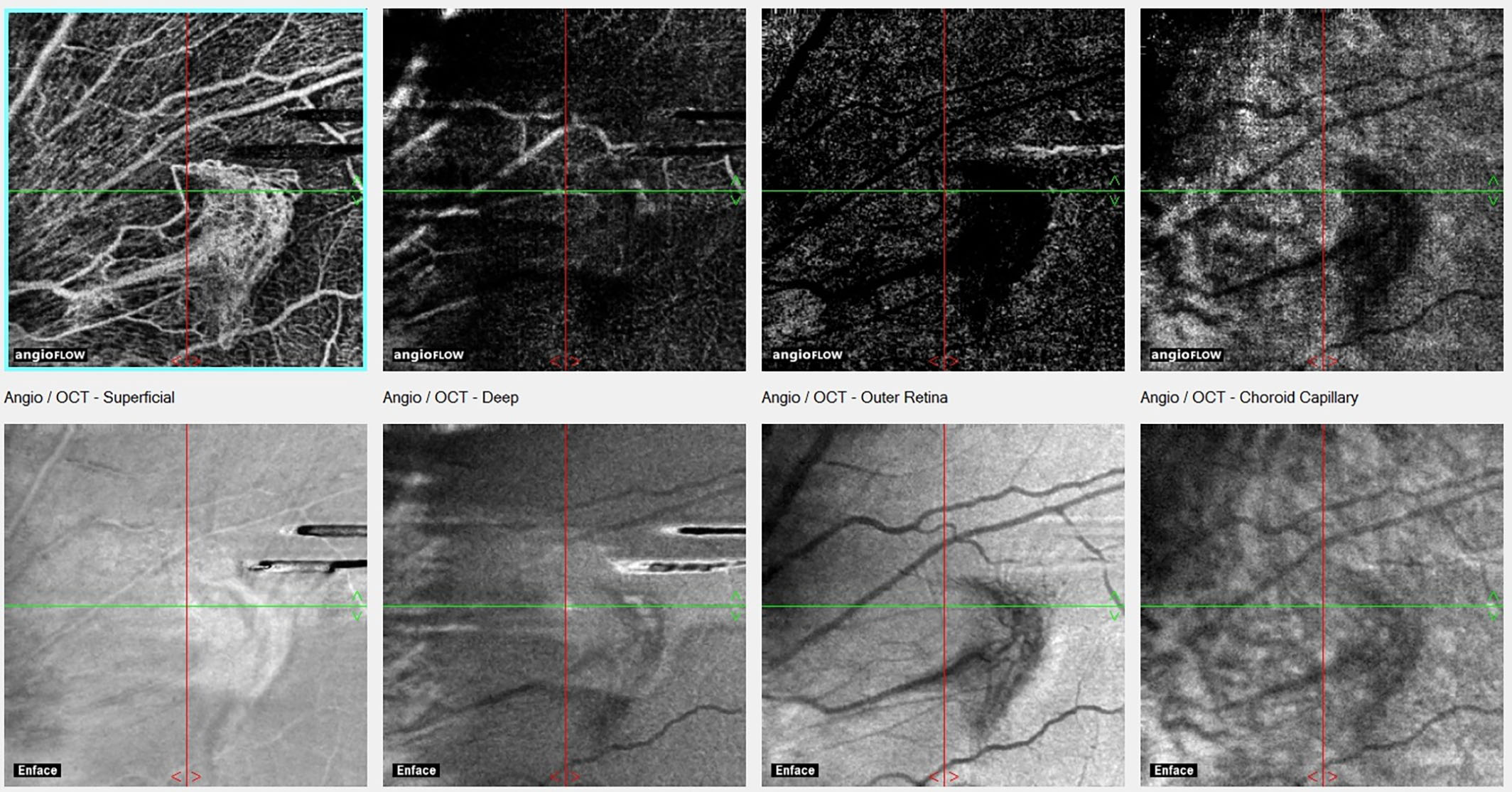
What technique has been used to produce these images (below) and what might they reveal?
Case 11

How has this image (left) been captured and what advantages might this technique have over standard digital photography of the retina.
Case 12
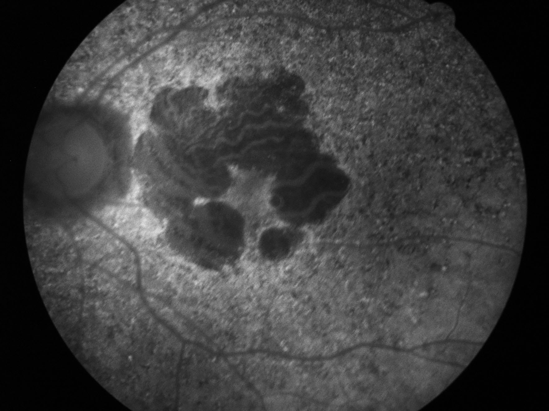
What technique is used to produce this image? What is the significance of the dark areas and also the brighter areas immediately surrounding them?
- A full discussion of the answers will be published in two months’ time.
- The latest patient information leaflet about age-related macular degeneration from the College of Optometrists is free for College members to download at www.college-optometrists.org.