Domains and learning outcomes (C110034)
• One distance learning CPD point for optometrists.
Professionalism
Upon completion of this CPD, ECPs will be able to identify ocular signs of possible abuse as they relate to shaken baby syndrome (abusive head trauma) (s.11)
Upon completion of this CPD, ECPs will be able describe how to keep adequate notes on discovery of retinal haemorrhages (s.11)
Clinical practice
Upon completion of this CPD, ECPs will be able to list other causes of retinal bleeding which could be confused with shaken baby syndrome (s.5)
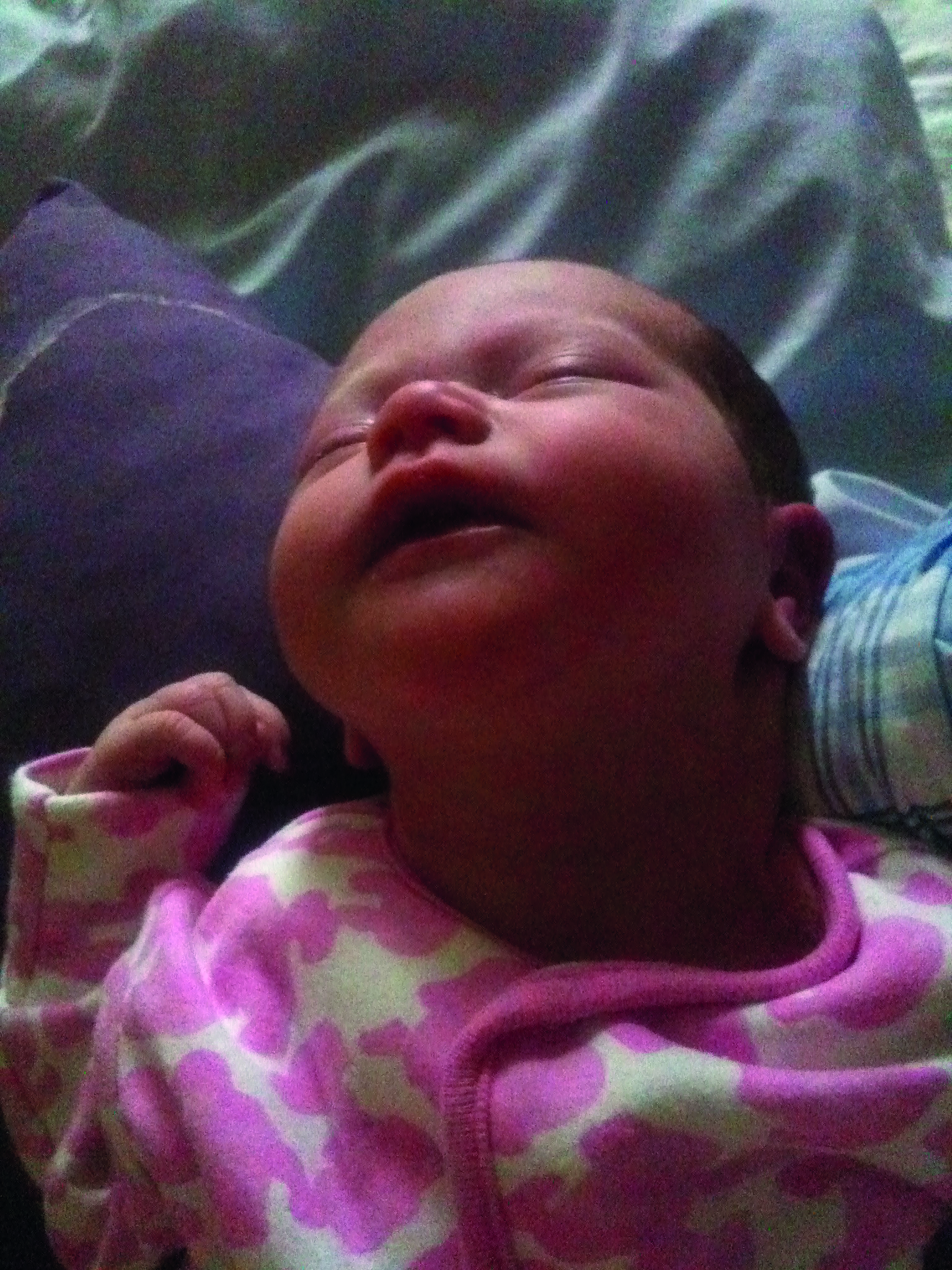
Abusive head trauma or shaken baby syndrome (SBS) is a brain injury thought to be caused by violent or excessive shaking of a baby. Although most commonly diagnosed in infants under the age of two it has been diagnosed in children as old as five.1 The triad of symptoms that are thought to indicate the presence of SBS are; brain swelling, retinal haemorrhages and subdural haematoma. SBS was first proposed by Norman Guthkelch, a neurosurgeon in Hull Royal Infirmary.
In a paper published in the British Medical Journal in 1971, he noted that although subdural haemorrhage was one of the most common findings in battered infants, many of these infants had no signs of external injury.2 Thus, he hypothesised that the shaking of an infant could cause these haemorrhages even in the absence of the child having been struck.
This hypothesis was further supported by a 1972 paper written by American paediatrician and radiologist John Caffey in which he suggested that ‘whiplash-shaking’ of an infant is a common cause of skeletal and brain injuries.3 His findings were based on a literature review of 27 cases of suspected child abuse. However, 15 of the cases were attributed to a single infant-nurse who admitted to shaking infants in her care. The case of the infant-nurse was found in a Newsweek article from 1956. Some of the other cases had evidence of, or a history of, external trauma.
In 1986, a literature review on SBS was published by Lucinda Dykes.4 In her review she notes that many questions remain on SBS, but she ultimately endorses recommendations made by Caffey regarding the routine fundus examination of infants and a public health campaign to warn people of the dangers of shaking babies. In all the literature reviewed by Dykes, there does not appear to be any cases where the author of a paper could state categorically that injuries were caused by shaking in the absence of any other trauma. There is a lot of ‘thought to be...’ and ‘might be...’ in these papers.
Guthkelch’s hypothesis was widely accepted in the early 70s but at the time of publication his hypothesis was not scientifically tested. In 1987, a paper was published in which the authors retrospectively examined 48 cases of infants and children diagnosed with SBS.5 Thirteen cases were fatal and the autopsies of all 13 cases showed evidence of blunt impact to the head that was not noted at the time of the SBS diagnosis.
In only one of the 48 cases was the child assumed to have been injured by shaking alone. The authors also made a whole infant head model in which they tested if shaking could produce the injuries associated with SBS. The authors concluded that the SBS injuries could only occur with some form of blunt trauma. However, their infant head model was later challenged.
In spite of questions around whether or not an infant could show the triad of symptoms from shaking alone, the American Academy of Paediatrics’ committee on child abuse and neglect published a paper in 2009 in which the authors recommended that the term ‘shaken baby syndrome’ should be replaced with ‘abusive head trauma’ to illustrate the fact that the triad of symptoms associated with SBS can also be caused by other forms of head trauma, notably by blunt impact.6
The committee still maintain that shaking alone can cause these symptoms. Now even the term ‘abusive head trauma’ is no longer considered appropriate, as to call the injuries by this name, implies that the medical professional knows that the injuries were caused by abuse, as opposed to by accident. The preferred term is now ‘retinodural haemorrhage of infancy’.
In a 2023 review of evidence for SBS the authors concluded: ‘Research has shown the triad is not sufficient to infer shaking or abuse and the shaking hypothesis does not meet the standards of evidence-based medicine.’7 This paper by Waney Squier, a neuropathologist who specialises in the foetal and neonatal brain, was almost immediately followed up by a comment from Lori Frasier, a medical expert on child abuse and neglect, refuting Squier’s findings.8 The same issue of the journal contained a letter from SBS expert Chris Brook in support of Squier’s findings.9
Where does all this controversy leave the eye care professional (ECP) when faced with an infant with unexplained retinal bleeding? What is the typical presentation of the retinal haemorrhages and what else might cause retinal bleeding in an infant?
It is unlikely that an ECP would be the first healthcare professional to see a child under the age of five with the signs of SBS, as often the infants are visibly unwell and in view of their young age, the parents normally take their child to a doctor or hospital in the first instance. Patients may present with lethargy, problems with feeding and irritability.
The retinal haemorrhages seen in SBS are thought to be caused by movement of the vitreous, which in turn places traction on the retina. The haemorrhages are generally bilateral, but they can be unilateral.10 (Although the mechanism by which a shaken baby only has unilateral haemorrhages appears to not be fully understood). The haemorrhages are normally multiple and they extend out to the ora serrata.
Depending on the severity of the injury the haemorrhages may be pre-retinal (ie in the vitreous), intra-retinal or subretinal or they may be multilayered.10 Intraretinal haemorrhages may be dot or blot shaped if they are in the inner nuclear and outer plexiform layer of the retina. Haemorrhages in the nerve fibre layer are flame-shaped and may contain Roth’s spots.11, 12 These are white patches in the centre of retinal nerve fibre layer haemorrhages. Subretinal haemorrhages can occur as a result of bleeding from the retina and/or choroid. These may be located above the retinal pigment epithelium (RPE) or below, known as sub-RPE haemorrhages. Because of their depth, these haemorrhages are less bright than intraretinal haemorrhages.
The vitreous haemorrhages are postulated to arise as a result of retinal haemorrhages slowly breaking through to the vitreous and therefore, they may not be present until a few days after the initial injury. An observational case series of five infants with suspected SBS showed that the retinal haemorrhages resolved fully within two to two-and-a-half weeks of injury.13
A 2023 systematic review found the presence of retinoschisis and retinal folds is 100% predictive of SBS in the absence of history of any other trauma.14 Retinoschisis is the splitting of the retinal layers. In SBS, it often occurs under the macula (but can occur elsewhere). The pre-macular cavity fills with pooled blood from the retinal haemorrhages. The pooled blood commonly forms a boat shape.15 The retinal fold is most often seen in a ring shape around the macula.
Other possible causes of retinal bleeding that could be confused with SBS are relatively rare. Short falls by infants that were witnessed were found to cause the triad of SBS including retinal haemorrhages.16 In a 2023 study in Japan, 32 infants and toddlers who were found to have had short falls that were deemed to be accidental, had a clinical finding of retinal haemorrhage.17
Case study
- A 13-month-old infant (Baby V) had a short fall from a sofa onto a marble floor. Baby V looked stunned initially and then started to cry. When the mother lifted baby V, she felt that the infant’s head was at an odd angle and so she called an ambulance. When the paramedics arrived, they checked baby V for broken bones and assured the mother that the infant was fine, but the mother was insistent that there was a problem and so the paramedics took the mother and infant to hospital. After more checks for broken bones at the hospital the infant was discharged. At 5am the following morning the mother woke and saw that baby V looked like she had had a stroke with a weakness all down her right side. After several days of testing in hospital baby V was diagnosed as having had a basal ganglia infarct. She had not had any infections and there was no family history that might explain why this had happened. An ophthalmologist confirmed that there was no ocular involvement. Baby V is now six years of age and fortunately has had no long-term effects from her injury. Basal ganglia infarction is a rare but known complication of minor head trauma in children.
Patients may present with lethargy
Other causes
- A 2006 paper used computer modelling to show that retinal bleeding could be caused by paroxysmal coughing, ie a coughing fit that is uncontrollable and violent.18 However, in 2017 researchers tested this hypothesis by carrying out a dilated retinal examination on infants hospitalised with whooping cough in New Zealand. Forty-eight infants with paroxysmal coughing (all of whom were still coughing at the time of examination) were examined and no retinal haemorrhages were seen.19
- Ehlers Danlos syndrome (EDS) is an inherited connective tissue disorder. Two of the EDS subtypes have been associated with vitreous and retinal haemorrhages.20 The two subtypes are vascular EDS and musculocontractural EDS. They are both very rare subtypes with musculocontractural EDS only having been reported in fewer than 100 patients worldwide.
- Von Willebrand disease is an inherited bleeding disorder that often goes undiagnosed, as signs of the disease can be very mild. It has been shown to be associated with vitreous and retinal haemorrhages.21, 22 It is thought to affect about 1% of the population.
- Retinal and vitreous haemorrhages can occur with leukaemia and in rare cases these haemorrhages have been seen in the initial presentation of the disease prior to diagnosis.23 Roth spots can sometimes be seen in haemorrhages associated with leukaemia.
- Cerebral venous sinus thrombosis is when a blood clot forms in the brain’s venous sinuses. The clot may prevent blood from draining from the brain and this can cause vessels to swell and burst. In a 2022 paper, researchers suggest that this condition can cause subdural and retinal haemorrhages, which are likely to be misdiagnosed as SBS.24 The prevalence of this condition in those aged under 18 is one in 100,000.
- Retinal haemorrhages can be caused by coagulation and haematological disorders (including disseminated intravascular coagulopathy, haemophilia, idiopathic thrombocytopenic purpura), infection (particularly meningitis), metabolic disorders such as glutaric acidurias, and galactosemia. However, retinal haemorrhage very rarely presents in these conditions pre-diagnosis. In a 2021 review of retinal haemorrhage and bleeding disorders in children, the authors found 61 articles representing 32 children who were less than five years old and had retinal haemorrhage and a bleeding disorder.25 Of these 32 cases, the authors felt that there were only five possible cases where the retinal haemorrhage could have been mistaken for a sign of SBS. These cases included haemophilia, disseminated intravascular coagulation and von Willebrand disease. Disseminated intravascular coagulopathy is a blood clotting disorder that can be caused by infection or injury.
On discovery of retinal haemorrhages in a young child, ECPs should take a very careful history from the parent or carer. An explanation that is vague or inconsistent with medical findings is considered a possible flag for child abuse. The infant with retinal haemorrhages should be urgently referred for further medical evaluation, in order to check for cranial haemorrhages.
A follow-up call to the hospital should be made to ensure that the child has been seen. The child’s general practitioner should also be notified. Any referral letter should only contain the facts, ie a precise description of the clinical findings or images and a relaying of the history of any possible event that may explain the injury as told by the parent or carer. It is important to avoid any speculation in reporting the history. The finding of retinal haemorrhages alone is not sufficient to make a report of possible child abuse and it is not within the remit of an ECP to check a child for bruises. However, it is within our duty of care to ensure that the child is seen by another medical professional.
- Claire McDonnell is an optometrist and lecturer at Technological University Dublin. She is also a member of BUCCLE (British and Irish University and College Contact Lens Educators). Her research area is specialist contact lenses. She has presented in the UK, Ireland and Europe on contact lenses and optometric education.
- DOCET and ABDO offer safeguarding training for ECPs.
References
- Shaken baby syndrome. ADAM medical encyclopedia. Last reviewed by Kaneshiro, NK, Dugdale, DC, Conaway, B on 24/01/2023. Available at https://medlineplus.gov/ency/article/007578.htm Accessed on 09/08/2024
- Guthkelch AN. Infantile subdural haematoma and its relationship to whiplash injuries. Br Med J. 1971 May 22;2(5759):430-1.
- Caffey J. On the theory and practice of shaking infants. Its potential residual effects of permanent brain damage and mental retardation. Am J Dis Child. 1972 Aug;124(2):161-9
- Dykes LJ. The whiplash shaken infant syndrome: what has been learned? Child Abuse Negl. 1986;10(2):211-21.
- Duhaime AC, Gennarelli TA, Thibault LE, Bruce DA, Margulies SS, Wiser R. The shaken baby syndrome. A clinical, pathological, and biomechanical study. J Neurosurg. 1987 Mar;66(3):409-15.
- Christian CW, Block R; Committee on Child Abuse and Neglect; American Academy of Pediatrics. Abusive head trauma in infants and children. Pediatrics. 2009 May;123(5):1409-11
- Squier W. Retinodural haemorrhage of infancy, abusive head trauma, shaken baby syndrome: The continuing quest for evidence. Dev Med Child Neurol. 2024 Mar;66(3):290-297.
- Frasier L. Retinodural haemorrhage of infancy: Support for continuing current terminology and approaches. Dev Med Child Neurol. 2024 Mar;66(3):273-274.
- Brook CB. The evidence for shaken baby syndrome (abusive head trauma) is still flawed. Dev Med Child Neurol. 2024 Mar;66(3):398-399.
- Morad Y, Kim YM, Armstrong DC, Huyer D, Mian M, Levin AV. Correlation between retinal abnormalities and intracranial abnormalities in the shaken baby syndrome. Am J Ophthalmol. 2002 Sep;134(3):354-9.
- Ruddy SM, Bergstrom R, Tivakaran VS. Roth Spots. [Updated 2023 Jul 17]. In: StatPearls [Internet]. Treasure Island (FL): StatPearls Publishing; 2024 Jan-. Available from: https://www.ncbi.nlm.nih.gov/books/NBK482446/ Accessed 14th Aug 2024.
- Pittner, A, Philips, H, Patel, S, Kolata, I. Roth Spots. [Updated 2024 Jan 19]. In: EyeWiki [Internet]. American Academy of Ophthalmology. Available from: https://eyewiki.org/Roth_Spots Accessed 14th Aug 2024.
- Esposito, DM, Nguyen, TL, Josephberg, RG. The medical-legal implications of rapid clearance of retinal haemorrhaging in shaken baby syndrome (SBS). ARVO annual meeting abstract. Invest Ophthalmol Vis Sci. April 2009, Vol.50, 3181.
- Di Fazio N, Delogu G, Morena D, Cipolloni L, Scopetti M, Mazzilli S, Frati P, Fineschi V. New Insights into the Diagnosis and Age Determination of Retinal Hemorrhages from Abusive Head Trauma: A Systematic Review. Diagnostics (Basel). 2023 May 12;13(10):1722.
- Greenwald MJ, Weiss A, Oesterle CS, Friendly DS. Traumatic retinoschisis in battered babies. Ophthalmology. 1986 May;93(5):618-25.
- Plunkett J. Fatal pediatric head injuries caused by short-distance falls. Am J Forensic Med Pathol. 2001 Mar;22(1):1-12.
- Kato M, Nonaka M, Akutsu N, Narisawa A, Harada A, Park YS. Correlations of intracranial pathology and cause of head injury with retinal hemorrhage in infants and toddlers: A multicenter, retrospective study by the J-HITs (Japanese Head injury of Infants and Toddlers study) group. PLoS One. 2023 Mar 17;18(3):e0283297.
- Geddes JF, Talbert DG. Paroxysmal coughing, subdural and retinal bleeding: a computer modelling approach. Neuropathol Appl Neurobiol. 2006 Dec;32(6):625-34.
- Raoof N, Pereira S, Dai S, Neutze J, Grant CC, Kelly P. Retinal haemorrhage in infants with pertussis. Arch Dis Child. 2017 Dec;102(12):1158-1160.
- Asanad S, Bayomi M, Brown D, Buzzard J, Lai E, Ling C, Miglani T, Mohammed T, Tsai J, Uddin O, Singman E. Ehlers-Danlos syndromes and their manifestations in the visual system. Front Med (Lausanne). 2022 Sep 27;9:996458.
- Herrmann WA, Lohmann CP, Demmler-Hackenberg M, Gabel VP. Von Willebrand’s disease type I as a cause for subvitreal, retinal and subretinal haemorrhages. Graefes Arch Clin Exp Ophthalmol. 2005 Apr;243(4):383-5.
- Shiono T, Abe S, Watabe T, Noro M, Tamai M, Akutsu Y, Ishikawa M, Suzuki S, Mori K. Vitreous, retinal and subretinal hemorrhages associated with von Willebrand’s syndrome. Graefes Arch Clin Exp Ophthalmol. 1992;230(5):496-7.
- Khurana M, Singh A, Pal H, Gupta AK, Kumar B. Ocular involvement-An unusual initial presentation of chronic myeloid leukemia: A case report. J Family Med Prim Care. 2023 Jul;12(7):1460-1463.
- Vaslow DF. Chronic subdural hemorrhage predisposes to development of cerebral venous thrombosis and associated retinal hemorrhages and subdural rebleeds in infants. Neuroradiol J. 2022 Feb;35(1):53-66.
- Thau A, Saffren B, Zakrzewski H, Anderst JD, Carpenter SL, Levin A. Retinal hemorrhage and bleeding disorders in children: A review. Child Abuse Negl. 2021 Feb;112:104901.