Being able to capture with ease high resolution images of the retina of adequate field using a portable device has long been wished for by many a practitioner. The strong association between eye health and age, the changing UK demographic profile and the increasing numbers of elderly who are housebound or have mobility problems mean this is not a selfish demand. Added to this, the ability to complement desktop cameras in a busy practice with one that can be carried between consulting rooms makes a good portable camera yet more attractive.
Over the last decade I have tried many a portable system, and a significant improvement in each subsequent design has been apparent. Resolution has improved, systems have become lighter, image transfer has developed to embrace wireless technology, and supporting software has allowed images to be manipulated or enhanced to levels that are clinically more than acceptable. One main hurdle has always been ease of use, with too many images out of focus or significantly contracted by shadowing or reflections. I recently got hold of the new Optomed Smartscope PRO (UK distributor Mainline Instruments) and, with these expectations in mind, tried it out.
The Smartscope PRO
The Smartscope PRO is a hand held imaging device with a five megapixel sensor and 2GB storage capability (figure 1). The unit comprises an operating handle and attachable retinal viewing unit (figures 2 and 3), anterior viewer (figure 4) and base unit, which both charges the handle and acts as a transfer dock for image upload to a computer (figure 5). I also had access to the optional slit lamp mount (figure 6), useful for steadying the unit for better accuracy. At just 400g weight and with 90 minutes’ continual use when fully charged, the Smartscope PRO easily meets the hardware requirements for a portable camera.
The unit comes with useful Optomed Workstation software (figure 7) which makes image selection, enhancement and storage with patient data easy, though equally images can simply be directly transferred via file explorer into the usual computer operating system. The EyFi Mobi software allows you to set up the system so that image data can be seamlessly transferred to the computer over any available WiFi – something most useful in a practice setting.
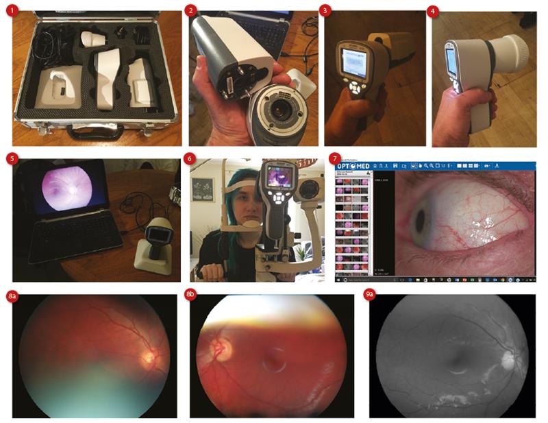
Image capture
I needed less time practising with the Smartscope PRO than previous cameras I had used before achieving satisfactory images. The system has an easily adjustable light setting, a moveable and variable brightness fixation target and options for manual and autofocusing. Unlike previous instruments of this sort, I found the autofocusing to be excellent. The unit can be programmed to take stills (colour, red free or infrared) or movie files, while the anterior viewer allows zoomed magnification of the ocular surface.
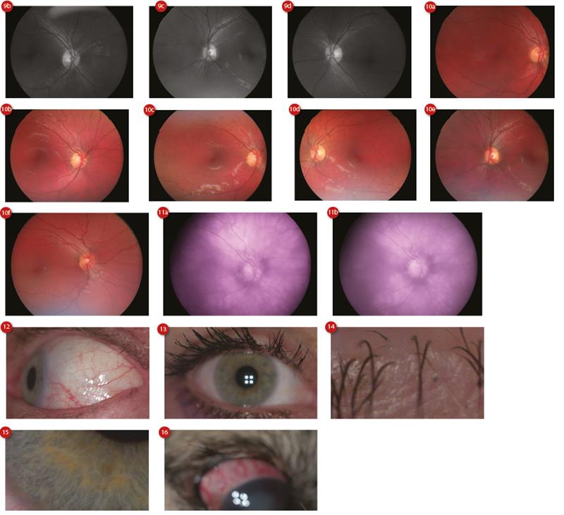
As always, alignment is essential during capture and, unless a green circle appears on screen to signify correct positioning, and axis adjustment has been undertaken to get rid of side encroachment upon the image, then image capture is best not continued. Figures 8a and b show the sort of images gained if this is not obeyed. I managed to operate the system on undilated patients with little difficulty and some of the results are displayed here; figure 9 showing red free, figure 10 colour and figure 11 infrared.
The resolution of anterior and ocular surface images was impressive as can be seen with the capture of bulbar hyperaemia (figure 12), lashes (figures 13 and 14) and limbal engorgement (figure 15). Perhaps a little too ambitious, I tried to image the cataracts of a small border terrier (figure 16) but its reluctance to behave meant the autofocus was just too slow.
I like this camera and suggest it is another step forward in portable imaging. Most of all, I was impressed with its easy operation.
Further information at www.main-line.co.uk