![]()
![]()


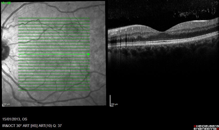
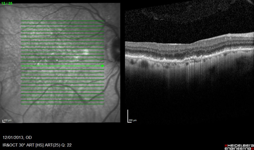
Identify the changes within the OCT image?
1. Marked retinal thickening and elevation.
2. Sub retinal fluid.
3. Sub retinal fibrosis.
4. RPE pigment migration.
5. Intra retinal oedema.
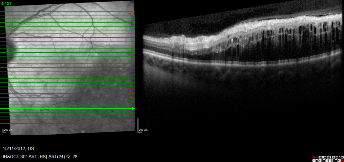
Sub retinal fluid
Sub retinal fibrosis
Pigment migration
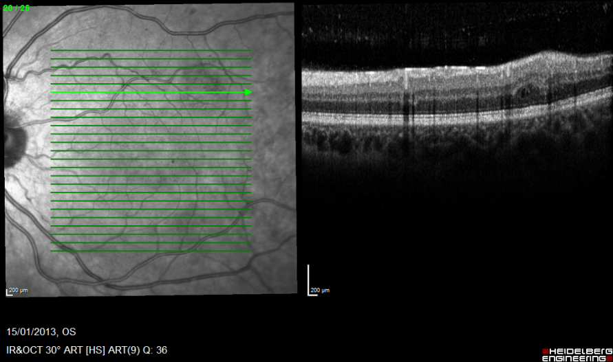
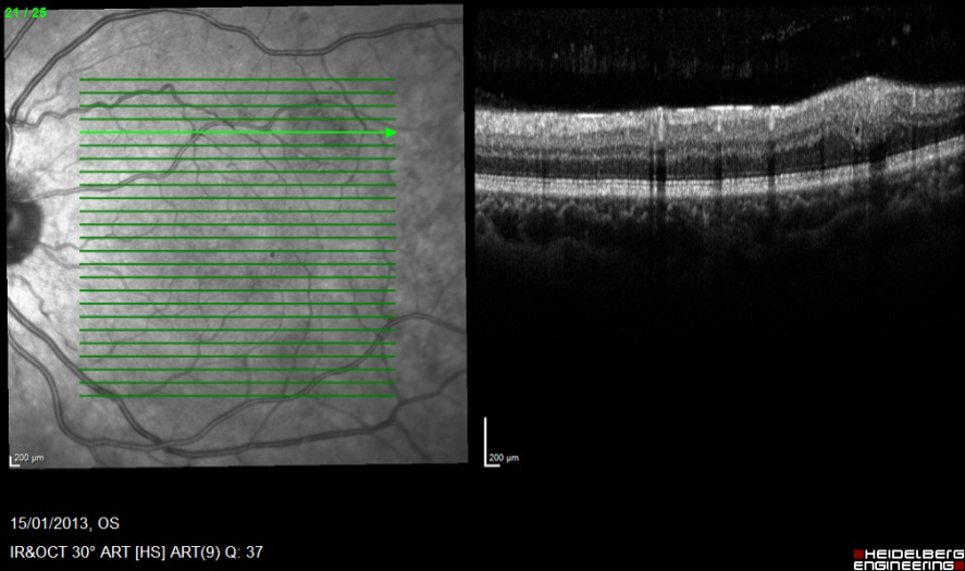
Assess the following OCT images of the same patient and identify the changes demonstrated within?
Inferior macular cross section
Superior macular cross section
Register now to continue reading
Thank you for visiting Optician Online. Register now to access up to 10 news and opinion articles a month.
Register
Already have an account? Sign in here