![]()
![]()


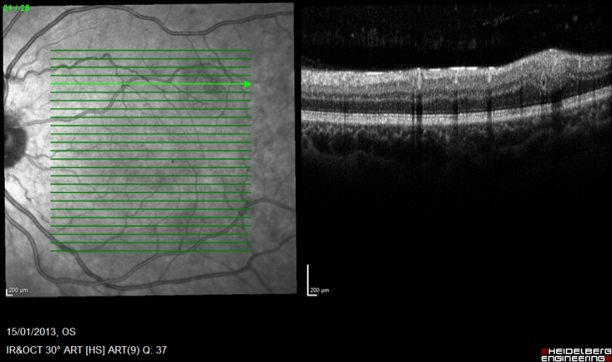
Foveal cross section
Section 1 is normal.
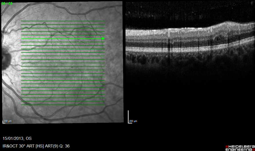
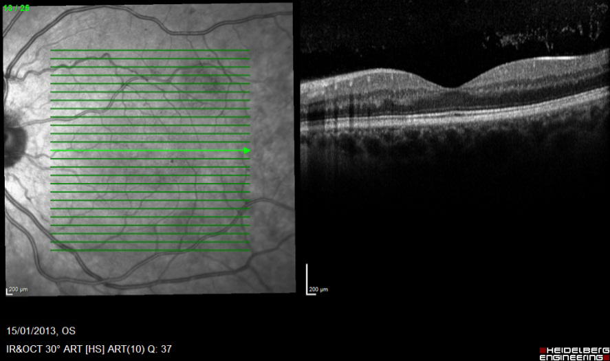
Superior macular cross sections.
Section 2 & 3, superior macular, show hypo reflective voids in the inner nuclear layer (3) and the outer nuclear layer (2) consistent with cystic oedema. Thickening of the retinal nerve fibre and ganglion cell layers, with increased reflectivity and shadowing of the outer retina.
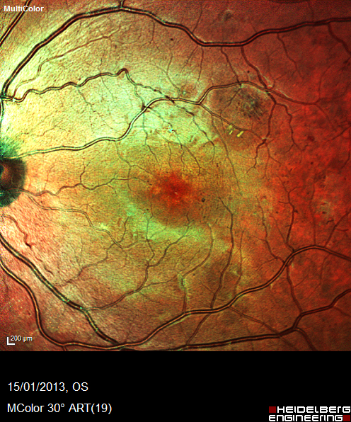
Describe the changes within the colour fundus image?
Register now to continue reading
Thank you for visiting Optician Online. Register now to access up to 10 news and opinion articles a month.
Register
Already have an account? Sign in here