![]()
![]()


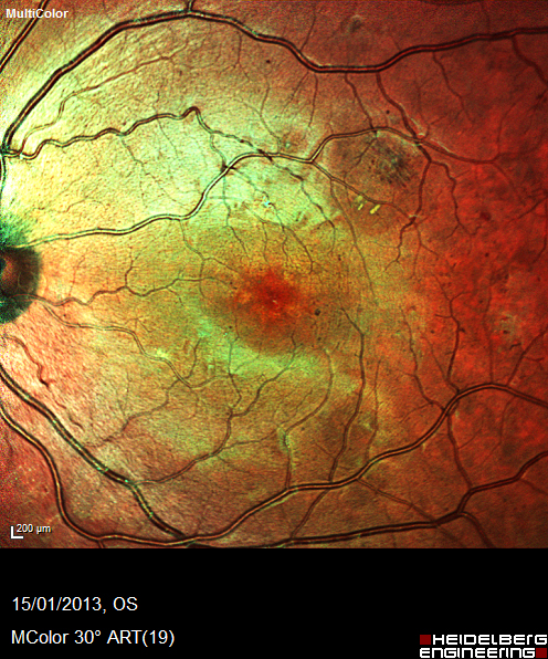
Colour fundus image:
1. Multiple red dots, microaneurysms and dot haemorrhages.
2. Retinal nerve fibre layer haemorrhage .
3. Exudate.
4. Small cotton wool spot (micro infarct) superior temporal.
5. Veins of normal calibre.
What would be your differential diagnosis?
Register now to continue reading
Thank you for visiting Optician Online. Register now to access up to 10 news and opinion articles a month.
Register
Already have an account? Sign in here