![]()
![]()


Describe the changes within the colour fundus image?
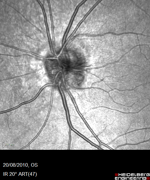
Colour fundus image:
1. Multiple red dots, microaneurysms and dot haemorrhages.
2. Retinal nerve fibre layer haemorrhage .
3. Extensive retinal nerve fibre layer oedema.
4. Cotton wool spot (micro infarct) inferior temporal.
5. Dilated veins of irregular calibre.
6. Macular oedema.
7. All changes are confined to one quadrant.
What would be your differential diagnosis?
1. Branch retinal vein occlusion
2. Hemisphere vein occlusion
3. Diabetic retinopathy
4. Hypertensive retinopathy
Register now to continue reading
Thank you for visiting Optician Online. Register now to access up to 10 news and opinion articles a month.
Register
Already have an account? Sign in here