![]()
![]()


What additional tests would you consider?
Monochromatic fundus imaging can be useful to assess the extent of retinal oedema, haemorrhage and microvascular change.
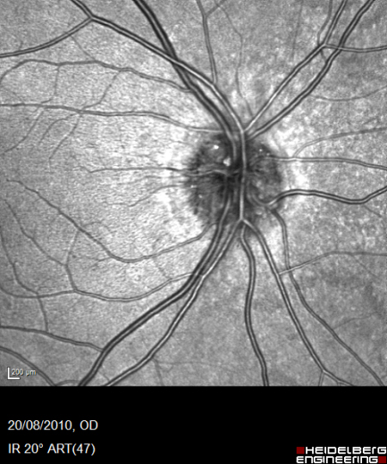
IR – retinal oedema
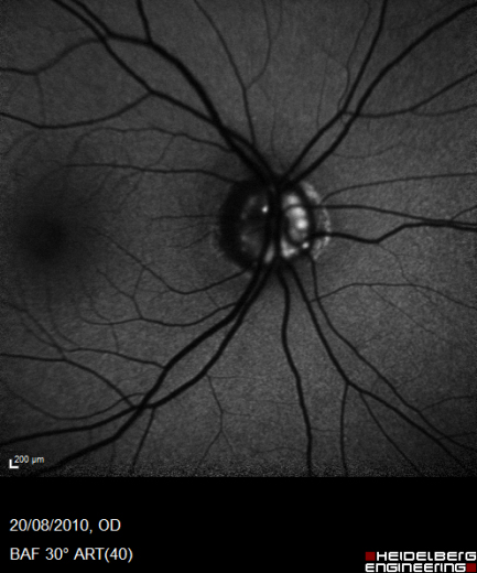
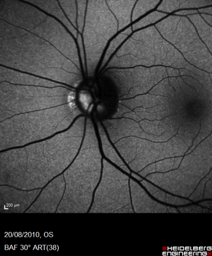
GR/BR – haemorrhage/microvascular change
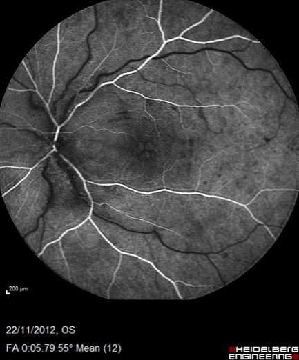
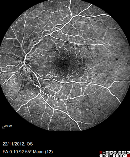
Additional tests performed in hospital might include fluorescein angiography to assess the extent of non-perfusion, presence of neovascularization and disruption to the foveal arcade.
Would you observe, discharge or refer to hospital eye service?
Case 3 will appear next week and, after all four cases are published, you will be presented with some related questions. After completing these you will be asked to submit some information about your management and you will then be sent information about all four cases plus confirmation of your points.
If you have any queries or wish for any further information please contact William.harvey@rbi.co.uk or send a message on our CET forum.