GR/BR – haemorrhage/microvascular change and retinal cysts.
![]()
![]()


What additional tests would you consider?
Monochromatic fundus imaging can be useful to assess the extent of retinal oedema, haemorrhage and microvascular change.
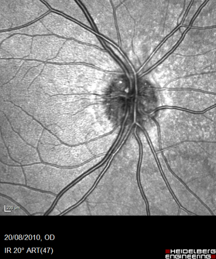
IR – retinal oedema
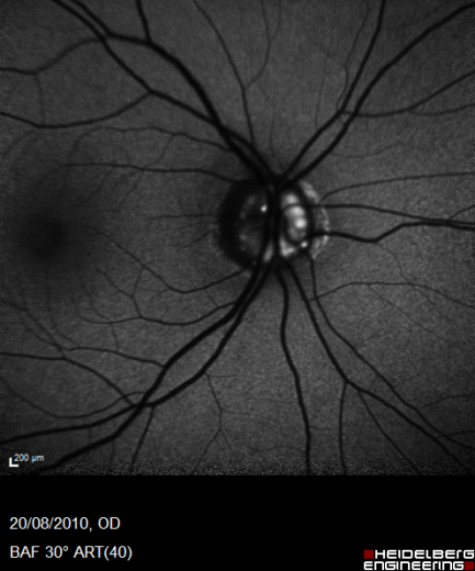

GR/BR – haemorrhage/microvascular change and retinal cysts
Additional tests performed in hospital might include fluorescein angiography to assess the extent of non-perfusion, presence of neovascularization and disruption to the foveal arcade.
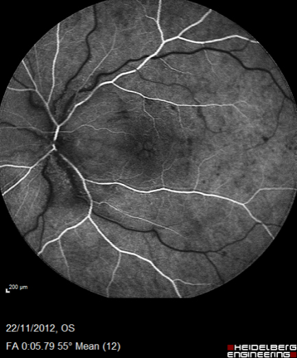
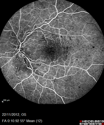

Fluorescein angiography shows delayed venous filling, mild non-perfusion and dilated capillaries, and disc, macular and nerve fibre layer oedema.
Register now to continue reading
Thank you for visiting Optician Online. Register now to access up to 10 news and opinion articles a month.
Register
Already have an account? Sign in here