![]()
![]()


Examine the images of the fellow eye and describe the changes?
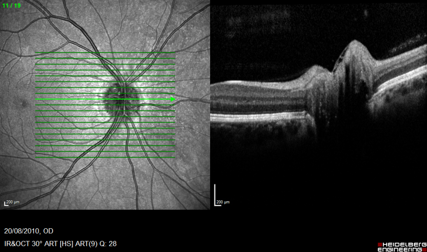
1. The elevation of the optic nerve head is less marked than in the left eye, there is a shallow cup and no evidence of retinal nerve fibre layer oedema. The neurosensory retinal structure is normal. The fundus image again shows anomalous arterial branching.
What additional tests might you recommend?
Register now to continue reading
Thank you for visiting Optician Online. Register now to access up to 10 news and opinion articles a month.
Register
Already have an account? Sign in here