Angioid streaks represent breaks in Bruch's membrane - the transparent, inner layer of the choroid situated adjacent to the retinal pigment epithelium. 
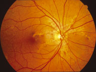
In patients with angioid streaks, the membrane is abnormally thickened, calcified and fragile. Approximately half of cases are idiopathic, and half occur in association with systemic disease:
Pseudoxanthoma elasticum (most common) - an inherited connective tissue disorder. Approximately 85 per cent of patients with pseudoxanthoma elasticum have angioid streaks. Patients develop loose skin ('chicken skin') and accelerated atherosclerosis, and have an increased risk of gastrointestinal bleeding
Paget's disease - a chronic, progressive disease resulting in abnormal, deformed, thickened and brittle bones. Features include bone pain, bowing of the legs, pathological fractures, hearing loss, enlargement of the skull and congestive cardiac failure. (Optic atrophy is a rare complication of Paget's disease, due to bony compression of the optic canal) Less common causes - other connective tissue disorders (such as Ehlers-Danlos syndrome and Marfan syndrome), haemoglobinopathies (for example, sickle cell disease), acromegaly and lead poisoning.
SYMPTOMS
Angioid streaks are often asymptomatic. Reduced vision or distorted shapes of objects (metamorphopsia) may occur through one or more of the following mechanisms:
Angioid streak traversing the fovea
Choroidal rupture with subfoveal haemorrhage, classically after relatively trivial ocular trauma
Neovascular vessels penetrating through breaks in Bruch's membrane. These friable, abnormal vessels may result in subretinal hemorrhage, retinal detachment, scarring and macular degeneration.
SIGNS
Clinical signs of an underlying disease may be present, and visual acuity may be reduced.
Angioid streaks are visible as jagged, irregular, dark red lines radiating from the vicinity of the optic disc and passing beneath retinal vessels. They often communicate in a ring around the optic disc. They are initially quite subtle, and become more obvious over time. Choroidal neovascular vessels or membranes may be visible.
The fundus characteristically assumes a mottled appearance (peau d'orange, particularly temporal to the macula). Other findings may include small white or salmon-coloured, peripheral chorioretinal scars ('histo-like spots') and optic disc drusen.
PREVALENCE
Rare.
SIGNIFICANCE
Often associated with systemic disease; may cause irreversible loss of acuity.
DIFFERENTIAL DIAGNOSIS
Myopic degeneration (lacquer cracks); choroidal rupture; choroidal folds.
SEE ALSO
Age-related macular degeneration; retinal detachment; optic disc drusen.
MANAGEMENT
Imaging investigations
Intravenous fluorescein angiography will confirm the presence of angioid streaks when the ophthalmoscopic appearance is subtle. The streaks are hyperfluorescent, and the retinal pigment epithelium is often irregularly clumped or mottled. Areas of neovascularisation leak fluorescein.
Blood tests
Consider when an underlying condition is suspected (for example, serum alkaline phosphatase and urinary calcium for Paget's disease; skin biopsy for pseudoxanthoma elasticum; haemoglobin electrophoresis for sickle cell anaemia). Referral to a physician is often appropriate for investigation and management of these conditions.
Laser
Treatment of choroidal neovascularisation associated with angioid streaks is controversial. Focal laser treatment has been applied with the objective of stabilising vision. Photodynamic therapy using verteporfin (see age-related macular degeneration) is an area of current research.
Advice
As minor trauma may cause rupture of the fragile Bruch's membrane with its attendant complications, patients are best advised to avoid contact sports and to wear safety glasses in situations that expose them to potential ocular trauma.
Review at six-month intervals is recommended, with particular attention to the presence or progression of neovascularisation. The patient may be supplied with an Amsler's grid for use at home, and advised to report any change.