Features

The Nidek AFC-210 doing the work for you

Instruments
Optician looks at the latest camera from Nidek and finds it boasts some useful features for the busy practitioner

Optician looks at the latest camera from Nidek and finds it boasts some useful features for the busy practitioner

p30NidekAFC-210.jpg
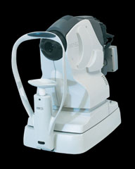
The first sight of the Nidek AFC-210 was at last year's Optrafair, where the prototype (the ANM-3000) created something of a stir. The incorporation of an autofocus facility was very attractive.

Any screening test ideally adheres to a number of basic principles. It should be accurate enough to detect any lesions relative to its function (so have a good sensitivity), as well as being specific enough to allow normal eyes to be seen as exactly that.

For any good retinal camera, this really is a matter of ensuring good image quality that can be easily interpreted. The new AFC-210 has several features which ensure just that.

Another recommendation is that the system should be easily operated, if necessary by non-professional staff, and the AFC-210 has features that make it almost impossible not to get an excellent quality image with any clear media patient.

Increased resolution

The final product displays significant modifications on the prototype instrument unveiled last year.

The original 3 megapixel resolution has been increased, so that the AFC-210 now boasts 12 megapixels, giving excellent resolution, even of enlarged images. As a Birmingham Optical Group technician noted: 'Three million was just not sufficient. We wanted to offer more than competitor instruments.'

The high resolution and fine gradation of the image means details can be visualised clearly with minimum flashlight required. This in turn reduces discomfort for patients and the risk of inner limiting membrane reflection, the veiling glare that often appears on the images of young healthy patients.

Auto-alignment

The most outstanding features of the new camera, however, are the auto-tracking and auto-focusing.

Most cameras rely on the operator centring the camera before the pupil of the patient, usually by using a screen shot of the pupil and then moving the camera until the edge of the pupil is then aligned with an on-screen marker of some nature.

The camera is then moved toward the patient and the fundus viewing lens either automatically drops in or is set in position before markers appear on the retinal image on screen. A degree of focusing and alignment is required before an image is ready for capture.

Patient movement or operator misinterpretation of the screen image can make for shots which are shadowed or out of focus.

For patients with less than dilated pupils and clear media, experience and skill is reflected in better quality images.

The auto-alignment feature of the AFC-210 allows easy pupil alignment. The auto-focus facility then automatically switches to the retinal shot which then automatically sets the instrument and takes the best quality image. The joystick is not used for this part of the capture.

To add to this, the unit also incorporates an 'auto-chinrest' feature, automatically adjusting the patient head height to give perfect alignment. All this allows the manufacturer to boast that the images taken 'are never out of focus'.

While in the anterior eye mode, the camera is capable of taking excellent quality anterior eye images, usually undertaken by slit-lamp attached cameras. This significantly increases the usefulness of the instrument.

Image storage

Image storage and manipulation is easy. The software for capture is all inclusive in the main camera unit so it can be used without hook-up to a laptop or desktop computer. Images may be directly sent to a data transfer system, such as a memory pen, allowing images taken during one session to be uploaded to a computer when convenient.

The system will easily fit within the same area as, for example, an auto-refractor, so could usefully be included in a pre-screening area in a busy practice. The manufacturer stresses that the large high contrast buttons and lever make operation in a dark room easy, even when not light-adapted.

Once the images are loaded, either directly or indirectly, on to the computer, the Navis-lite software allows easy ordering. There are also a number of other useful features. As expected, there is the ability to zoom in to select the colour or appearance of display - red-free, grey scale, colour reversal, contrast red/green/blue, gamma control to enhance darker images and so on.

A panoramic viewing function allows merging of images to give wider field views. This is especially useful in conditions where there may be multiple lesions spread over a large area and it is useful to know how they position themselves relative to one another.

A C/D ratio and disc HV feature allows for disc measurement which is useful, particularly for looking for changes to disc topography over a period of time.

For all those budding lecturers and authors, there is also the usual facility to add labels to or draw over an image. For those who acquire the stereo viewer (an optional extra), the camera can take two disparate shots of the disc (or any other area for that matter) which may be viewed using the special viewer to give a three dimensional image on the screen.

Optician will be giving the system a road test in the next month and our findings will be published later in the year.

For further information on the new AFC-210, please contact Birmingham Optical Group on 0121 442 5800.