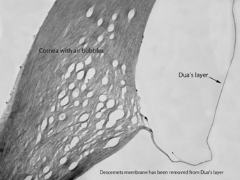
A sixth layer of the cornea has been identified and named after the scientist who discovered it.
The breakthrough, announced by The University of Nottingham last week, was expected to improve outcomes for patients undergoing corneal grafts and transplants.
Professor Harminder Dua, professor of ophthalmology and visual sciences at the university, had the extra layer named after him as the findings were published in Ophthalmology.
Dua's layer adds to the previously identified corneal epithelium, Bowman's layer, the corneal stroma, Descemet's membrane and the corneal endothelium.
Professor Dua said: 'This is a major discovery that will mean that ophthalmology textbooks will literally need to be re-written. Having identified this new and distinct layer deep in the tissue of the cornea, we can now exploit its presence to make operations much safer and simpler for patients.
Register now to continue reading
Thank you for visiting Optician Online. Register now to access up to 10 news and opinion articles a month.
Register
Already have an account? Sign in here