![]()
![]()


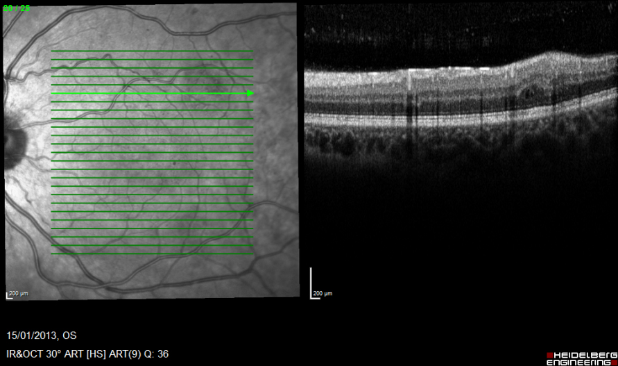
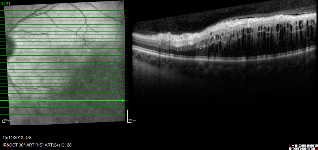
Colour fundus image:
Irregular subretinal pigmentation (red) at macular region. This pigmentary change extends from the optic nerve head toward the macular and radiates from the nerve into all quadrants. The reflected infrared (IR) fundus image (lower) confirms the changes observed within the colour image (upper), showing hyper reflective, sub retinal pigment.
What would be your differential diagnosis?
What additional tests might you recommend?
Register now to continue reading
Thank you for visiting Optician Online. Register now to access up to 10 news and opinion articles a month.
Register
Already have an account? Sign in here