Confirm your diagnosis?.
![]()
![]()


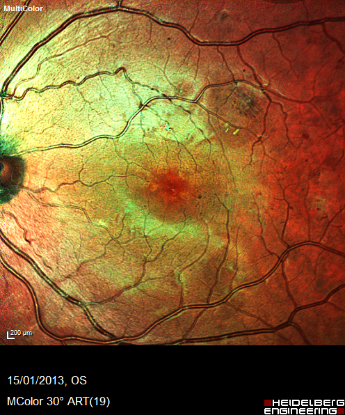
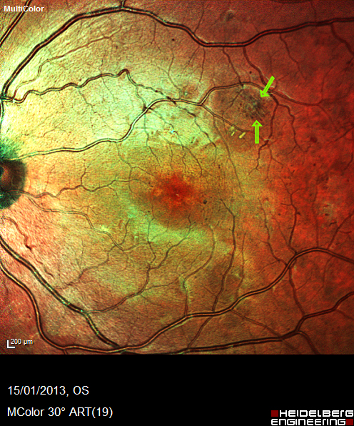
Fundus autofluorescence (FAF) imaging (Blue Peak) allows assessment of the integrity and metabolic activity within the RPE layer. Angioid streaks are a defect in Bruch’s membrane, these often appear as hypo-autofluorescent fissures (upper figure). Angioid streaks are often associated with retinal pigment epithelial atrophy (lower figure, red) which is preceded by hyper fluorescent patches (lower figure, green). Extension of the streaks towards the fovea has a poor prognosis.
Confirm your diagnosis?
Register now to continue reading
Thank you for visiting Optician Online. Register now to access up to 10 news and opinion articles a month.
Register
Already have an account? Sign in here
